Researchers explore an innovative method for single-cell analysis
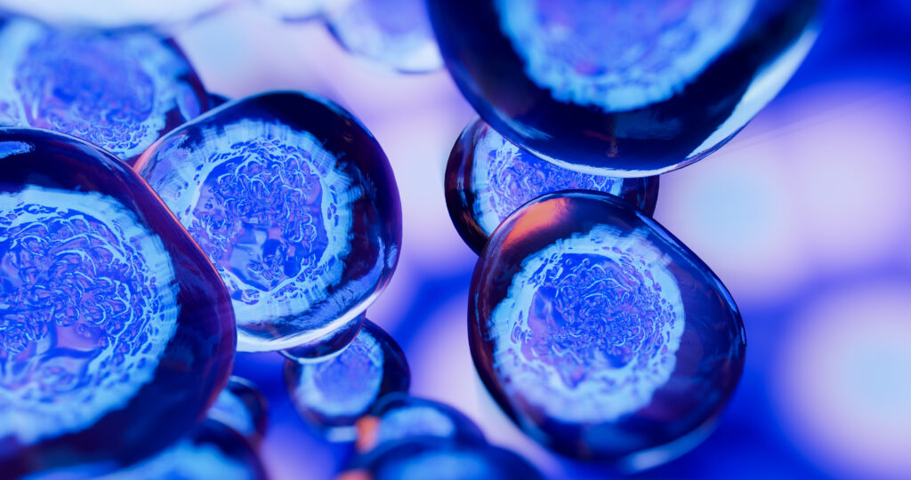
Cells produce proteins that serve different purposes in maintaining human health. These bioactive secretions range from growth factors to antibodies to cytokines and vary between different types of cells. Even within a certain cell type, however, there are individual cells that produce more secretions than others, a phenomenon that especially interests scientists studying cell-based therapies. In contrast to molecular therapies, which typically involve specific genes or proteins, a primary challenge to crafting cell therapies is the wide range of functional outputs seen in cells that have the same genetic template. This leads to the question of what molecular properties, from a genomic and transcriptomic perspective, would lead one cell to produce more of a protein than its companions.
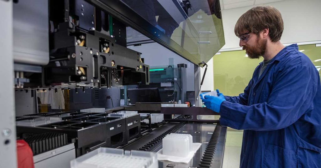
There have been few investigative strategies put forth that allow scientists to connect a cell’s characteristics and genetic coding with its secretions. In July 2023 a team of scientists published a paper in Nature Communications outlining an innovative solution: little hydrogel particles, or “nanovials”, that essentially serve as tiny test tubes and can be used to measure protein secretion, track transcriptome data, and identify relevant surface markers in a single cell.
Artificial intelligence (AI) is not a new technological development. The idea of intelligent machines has been popular for several centuries. The term “artificial intelligence” was coined by John McCarthy for a workshop at Dartmouth College in 1955 (1), and this workshop is considered the birthplace of AI research. Modern AI owes much of its existence to an earlier paper by Alan Turing (2), in which he proposed the famous Turing Test to determine whether a machine could exhibit intelligent behavior equivalent to—or indistinguishable from—that of a human.
The explosive growth in all things AI over the past few years has evoked strong reactions from the general public. At one end of the spectrum, some people fear AI and refuse to use it—even though they may have unwittingly been using a form of AI in their work for years. At the other extreme, advocates embrace all aspects of AI, regardless of potential ethical implications. Finding a middle ground is not always easy, but it’s the best path forward to take advantage of the improvements in efficiency that AI can bring, while still being cautious about widespread adoption. It’s worth noting that AI is a broad, general term that covers a wide range of technologies (see sidebar).
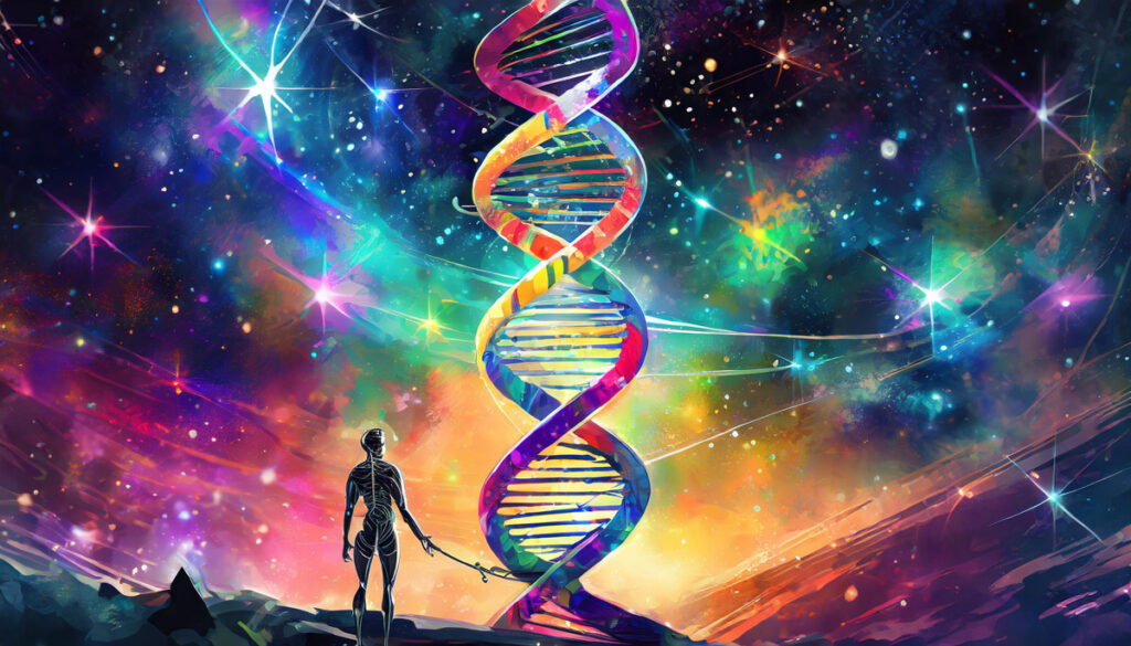
Image generated with Adobe Firefly v.2.
For life science researchers, AI has the potential to address many common challenges; a previous post on this blog discussed how AI can help develop a research proposal. AI can help with everyday tasks like literature searches, lab notebook management, and data analysis. It is already making strides on a larger scale in applications for lab automation, drug discovery and personalized medicine (reviewed in 3–5). Significant medical breakthroughs have resulted from AI-powered research, such as the discovery of novel antibiotic classes (6) and assessment of atherosclerotic plaques (7). A few examples of AI-driven tools and platforms covering various aspects of life science research are listed here.
Identifying Inflammasome Inhibitors: What’s Missing The NLRP3 inflammasome is implicated in a wide range of diseases. The ability to inhibit this protein complex could provide more precise, targeted relief to inflammatory disease sufferers than current broad-spectrum anti-inflammatory compounds, potentially without side effects.
Studies of NLRP3 inflammasome inhibitors have relied on cell-free assays using purified NLRP3. But cell-free assays cannot assess physical engagement of the inhibitor and target in the cellular micro-environment. Cell-free assays cannot show if an NLRP3 inhibitor enters the cell, binds the target and how long the inhibitor binding lasts.
Cell-based assays that interrogate the physical interaction of the NLRP3 target and inhibitor inside cells are needed.
One key obstacle to crafting effective gene therapies is the ability to tailor dosing according to a patient’s needs. This can be tricky because even if protein production is successful, staying within the therapeutic window is paramount—too much of a protein could be toxic, and too little will not produce the desired effect. This balance is difficult to achieve with current technologies. In a study recently published in Nature Biotechnology, researchers at Baylor College of Medicine investigated a possible solution to this problem, engineering a molecular “on/off” switch that could regulate gene expression and maintain protein production at dose-dependent, therapeutic levels.
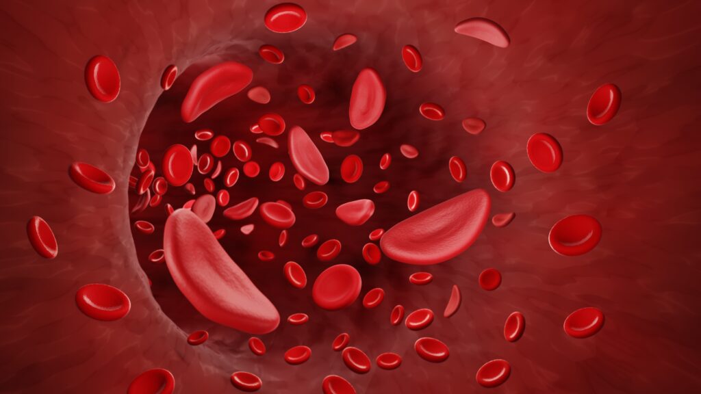
In sickle cell anemia, red bloods cells elongate into an abnormal “sickled” shape
Sickle cell disease is a debilitating blood disorder that causes recurrent pain crises and severe health effects, and can drastically impact quality of life. Recently, Vertex Pharmaceuticals and CRISPR Therapeutics introduced Casgevy, or exa-cel, a novel form of gene therapy that could radically change the management of sickle cell disease. On the heels of exa-cel’s approval in Britain, this groundbreaking therapy was also recently approved in the U.S.
Image adapted from original artwork by iSO-FORM LLC.
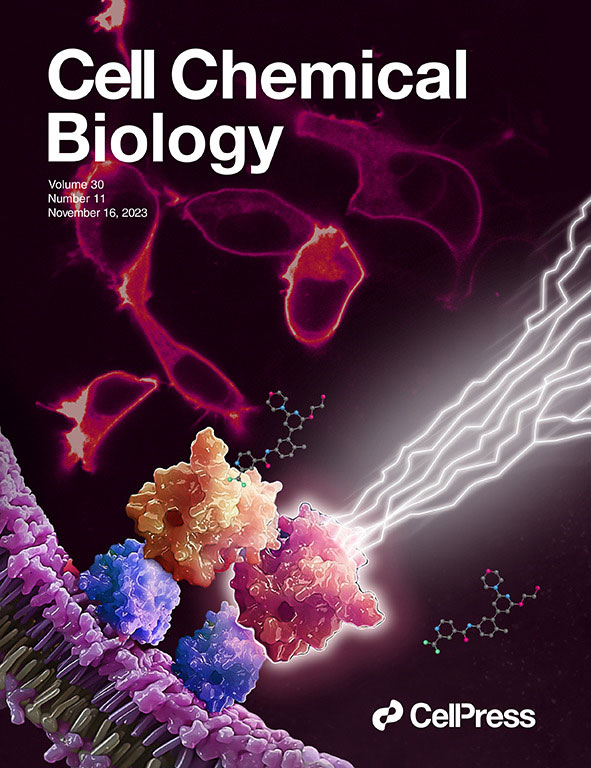
We made the cover! Of Cell Chemical Biology, that is.
This July, Cell Chemical Biology editors accepted a study from Promega scientists and invited the research team to submit cover art for the issue. The study in question details a BRET-based method to quantify drug-target occupancy within RAF-KRAS complexes in live cells. Promega scientists Matt Robers and Jim Vasta collaborated with one of our talented designers, Michael Stormberg, to craft an image that accurately represents the science in a dynamic and engaging way.
I spoke with Michael Stormberg to learn more about the creative process that went into creating this cover art and how he worked with the research team and other collaborators.
Imagination is often considered a uniquely human trait. Simply put, it is what allows us to think about things that aren’t happening in that moment, and it plays an integral part in our day-to-day lives. We use it when we think through our calendar for the day, consider restaurant options for dinner, or visualize the best route. It turns out this trait might not be as unique to humans as we thought. In fact, a study published in Science suggests that we might share this ability with rats (1).
Rats are the most divisive of rodents. Some people see disease-carrying scourges; some see intelligent, affectionate creatures with larger-than-life personalities; and still others simply can’t get past their bare tails and small eyes. Love them or hate them, science has shown that there is more to these creatures than meets the eye. They are intelligent, ticklish and empathetic; and the study in Science suggests, imaginative.
Recent research reveals that starfish anatomy is even stranger than previously thought
Most animals in the world are what biologists refer to as “bilateral”—their left and right sides mirror one another. It is also typically easy to tell which part of most animals is the top and which is the bottom. The anatomical arrangements of certain other animals, however, are slightly more confounding, for instance in the case of echinoderms, which include sea urchins, sand dollars and starfish. These animals are “pentaradial”, with five identical sections of the body radiating from a central axis. The question of how these creatures evolved into such a state has been a puzzle pondered by many a biologist, with little progress made until recently. In a new study published in Nature, scientists closely examining the genetic composition of starfish point to some key evidence that suggests a starfish is mostly just a head.
Starfish are a deuterostome, belonging to the superphylum Deuterostomia. Most deuterostomes are bilateral, leading scientists to believe that, despite their peculiar body plan, starfish evolved from a bilateral ancestor. This is supported by the fact that starfish larvae actually start out bilateral, and eventually transform into the characteristic star shape. But where the head of the starfish is, or whether it even has one, has proved difficult for scientists to parse out, especially since their outward structure offers no real clues.
There have been a number of theories posited, such as the duplication hypothesis—where each of the five sections of a starfish could be considered “bilateral”, placing the head at the center—and the stacking hypothesis, which asserts that the body is stacked atop the head. In a bilateral body plan, anterior genes broadly code for the front, or the head-region, and posterior genes are primarily responsible for the tail. The torso, or “trunk”, is the result of complex interplay between both anterior and posterior, as well as other types of genes. Researchers in this new study looked at the expression of these genes throughout the body plan as a possible source of clarity as to which part of the starfish is its head and which parts comprise the body.
To this end, researchers used advanced molecular and genetic sequencing techniques including RNA tomography and in situ hybridization. RNA tomography allowed them to create a three-dimensional map of gene expression throughout the limbs of the sea star Patiria miniate. In situ hybridization is a fluorescent staining technique that offered them a means by which to examine where exactly anterior or posterior genes are expressed in the sea star’s tissue, providing a clearer picture of genetic body patterning.
Remarkably, scientists found that anterior or head-coding genes were expressed in the starfish’s skin, including head-like regions appearing in the center, or midline, of each arm, while tail-coding genes were only seen at the outer edges of the arms. Perhaps even more remarkable was the lack of genetic patterning accounting for a trunk or torso, leading scientists to the conclusion that starfish are, for the most part, just heads.
Whether this holds true for other echinoderms remains to be proven, and further investigations into starfish anatomy may seek to pinpoint where in the timeline the trunk was lost. Overall, research like this helps scientists understand how life came to look the way it does. Oddly shaped creatures like the humble starfish can offer insight into the strange evolutionary processes that result in such rich biodiversity across the animal kingdom.
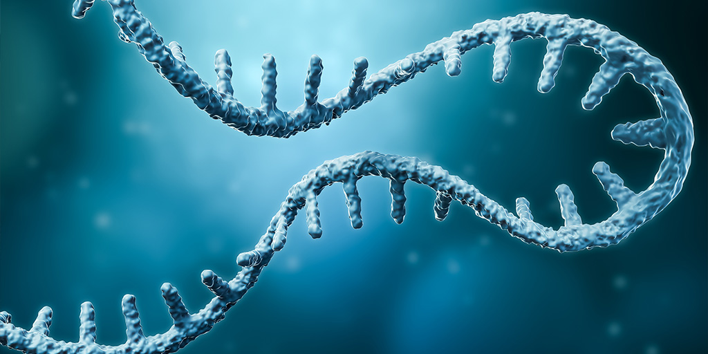
In recent years following the COVID-19 pandemic, RNA has gained attention for its successes and potential use in vaccines and therapeutics. One avenue of interest in RNA research is a non-coding class of RNA first identified almost 50 years ago, circular RNA (circRNA).
In 1976, Sanger et al. first identified circRNA in plant viroids, and later additions to the field found them in mice, humans, nematodes, and other groups. Unlike linear RNA, circRNA are covalently closed loops that don’t have a 5′ cap or 3′ polyadenylated tail. Following its discovery, researchers thought circRNA was the product of a rare splicing event caused by an error in mRNA formation leading to low interest in researching the subject (1).
In the early 2010s, following the development of high throughput RNA sequencing technology, Salzman et al. determined that circRNAs were not a result of misplicing, but a stable, conserved, and widely sourced form of RNA with biological importance. Since noncoding RNA makes up the majority of the transcriptome it’s an incredibly important field of study. We now recognize circRNAs for their potential as disease biomarkers and importance in researching human disease (2).
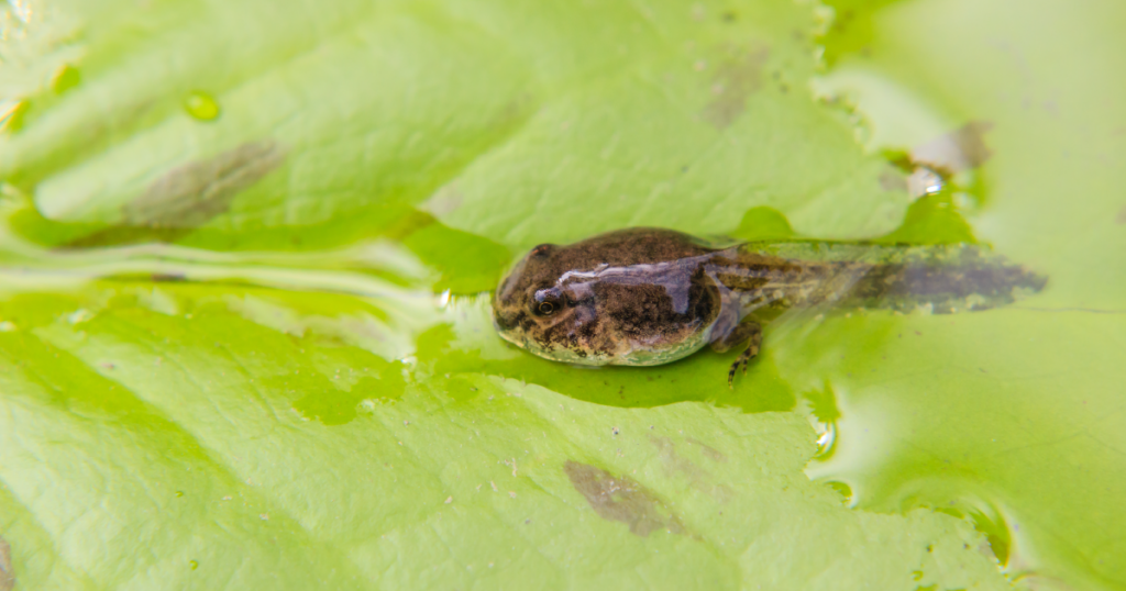
Amphibians are the most threatened vertebrate class worldwide. Because they lack the ability to regulate their own temperature and moisture levels, climate change is playing a significant role in this growing peril (1). Climate change impacts amphibian survival in several ways. In addition to habitat loss, growing drought conditions make maintaining body moisture levels challenging and warming temperatures restrict activity periods needed for reproduction as well as increasing the risk of heat stress.
Heat tolerance varies by species, and understanding what influences these differences could help predict species survival. The gut microbiota is known to affect a wide range of functions in host animals, and recently studies have begun to investigate its role in host thermal tolerance (2).
XWe use cookies and similar technologies to make our website work, run analytics, improve our website, and show you personalized content and advertising. Some of these cookies are essential for our website to work. For others, we won’t set them unless you accept them. To learn more about our approach to Privacy we invite you to Read More
By clicking “Accept All”, you consent to the use of ALL the cookies. However you may visit Cookie Settings to provide a controlled consent.
We use cookies and similar technologies to make our website work, run analytics, improve our website, and show you personalized content and advertising. Some of these cookies are essential for our website to work. For others, we won’t set them unless you accept them. To find out more about cookies and how to manage cookies, read our Cookie Policy.
If you are located in the EEA, the United Kingdom, or Switzerland, you can change your settings at any time by clicking Manage Cookie Consent in the footer of our website.
Necessary cookies are absolutely essential for the website to function properly. These cookies ensure basic functionalities and security features of the website, anonymously.
Cookie
Duration
Description
cookielawinfo-checbox-analytics
11 months
This cookie is set by GDPR Cookie Consent plugin. The cookie is used to store the user consent for the cookies in the category "Analytics".
cookielawinfo-checbox-functional
11 months
The cookie is set by GDPR cookie consent to record the user consent for the cookies in the category "Functional".
cookielawinfo-checbox-others
11 months
This cookie is set by GDPR Cookie Consent plugin. The cookie is used to store the user consent for the cookies in the category "Other.
cookielawinfo-checkbox-advertisement
1 year
The cookie is set by GDPR cookie consent to record the user consent for the cookies in the category "Advertisement".
cookielawinfo-checkbox-necessary
11 months
This cookie is set by GDPR Cookie Consent plugin. The cookies is used to store the user consent for the cookies in the category "Necessary".
cookielawinfo-checkbox-performance
11 months
This cookie is set by GDPR Cookie Consent plugin. The cookie is used to store the user consent for the cookies in the category "Performance".
gdpr_status
6 months 2 days
This cookie is set by the provider Media.net. This cookie is used to check the status whether the user has accepted the cookie consent box. It also helps in not showing the cookie consent box upon re-entry to the website.
lang
This cookie is used to store the language preferences of a user to serve up content in that stored language the next time user visit the website.
viewed_cookie_policy
11 months
The cookie is set by the GDPR Cookie Consent plugin and is used to store whether or not user has consented to the use of cookies. It does not store any personal data.
Analytical cookies are used to understand how visitors interact with the website. These cookies help provide information on metrics the number of visitors, bounce rate, traffic source, etc.
Cookie
Duration
Description
SC_ANALYTICS_GLOBAL_COOKIE
10 years
This cookie is associated with Sitecore content and personalization. This cookie is used to identify the repeat visit from a single user. Sitecore will send a persistent session cookie to the web client.
vuid
2 years
This domain of this cookie is owned by Vimeo. This cookie is used by vimeo to collect tracking information. It sets a unique ID to embed videos to the website.
WMF-Last-Access
1 month 18 hours 24 minutes
This cookie is used to calculate unique devices accessing the website.
_ga
2 years
This cookie is installed by Google Analytics. The cookie is used to calculate visitor, session, campaign data and keep track of site usage for the site's analytics report. The cookies store information anonymously and assign a randomly generated number to identify unique visitors.
_gid
1 day
This cookie is installed by Google Analytics. The cookie is used to store information of how visitors use a website and helps in creating an analytics report of how the website is doing. The data collected including the number visitors, the source where they have come from, and the pages visted in an anonymous form.
Advertisement cookies are used to provide visitors with relevant ads and marketing campaigns. These cookies track visitors across websites and collect information to provide customized ads.
Cookie
Duration
Description
IDE
1 year 24 days
Used by Google DoubleClick and stores information about how the user uses the website and any other advertisement before visiting the website. This is used to present users with ads that are relevant to them according to the user profile.
test_cookie
15 minutes
This cookie is set by doubleclick.net. The purpose of the cookie is to determine if the user's browser supports cookies.
VISITOR_INFO1_LIVE
5 months 27 days
This cookie is set by Youtube. Used to track the information of the embedded YouTube videos on a website.
Performance cookies are used to understand and analyze the key performance indexes of the website which helps in delivering a better user experience for the visitors.
Cookie
Duration
Description
YSC
session
This cookies is set by Youtube and is used to track the views of embedded videos.
_gat_UA-62336821-1
1 minute
This is a pattern type cookie set by Google Analytics, where the pattern element on the name contains the unique identity number of the account or website it relates to. It appears to be a variation of the _gat cookie which is used to limit the amount of data recorded by Google on high traffic volume websites.