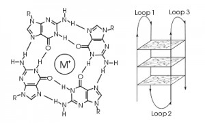
A guanine tetrad (left) and G-quadruplex (right). Image courtesy of Wikimedia Commons.
Proto-oncogenes are genes that organisms rely on for normal growth and development but, when mutated or dysregulated, can cause cells to grow uncontrollably, resulting in cancer and metastasis. In some cases, a single DNA mutation is sufficient for cancer to develop. Why then, do so many proto-oncogene promoters contain strings of guanine residues, which are extremely vulnerable to DNA damage from factors such as oxidative stress and hyperinflammation, to control transcription levels? From an evolutionary viewpoint, this is a contradiction: DNA sequences that are the most vulnerable to damage and mutation are key to regulating one of the cell’s most dangerous classes of genes. This seems to be a recipe for genomic instability and disease. Fortunately, evolution has provided a very clever solution to this potential problem.
Robert Hooke first coined the term “cell” after observing plant cell walls through a light microscope—little empty chambers, fixed in time and space. However, cells are anything but fixed.
Cells are dynamic: continually responding to a shifting context of time, environment, and signals from within and without. Interactions between the macromolecules within cells, including proteins, are ever changing—with complexes forming, breaking up, and reforming in new ways. These interactions provide a temporal and special framework for the work of the cell, controlling gene expression, protein production, growth, cell division and cell death.
Visualizing and measuring protein:protein interactions at the level of the cell without perturbing them is the goal of every cell biologist.
A recent article by Thomas Machleidt et al. published in ACS Chemical Biology, describes a new technology that brings us closer to being able to realize that goal.
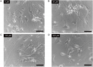
Ketamine induced morphological changes in neurons derived from iPSCs. Cells were treated with 0μM (Panel A), 20μM (Panel B), 100μM (Panel C) or 500μM (Panel D) ketamine for 24 hours. Scale bar = 50μm. From Ito, H., Uchida, T. and Makita, K. (2015) Ketamine causes mitochondrial dysfunction in human induced pluripotent stem cell-derived neurons. PLOS ONE10, e0128445. doi:10.1371/journal.pone.0128445.g002When I consider that major surgery was performed long before anesthetics were developed, I am grateful to be alive in the anesthesia era. Just the thought of being subjected to various cutting and retracting instruments without general anesthesia calls to mind a phrase: The cure is worse than the disease. Despite the advantages of unconsciousness during surgery, anesthesia can have side effects. Studies in neonatal nonhuman primates have demonstrated that the anesthetic ketamine has toxic effects. However, the differences between humans and nonhuman primates mean the outcome in one species is not the same in another. In an article recently published in PLOS ONE, scientists were interested in creating an experimental model of developing human neurons and using the model to better understand the toxic effects of ketamine on human cells. Continue reading “Developing a Model System to Test Ketamine Toxicity”
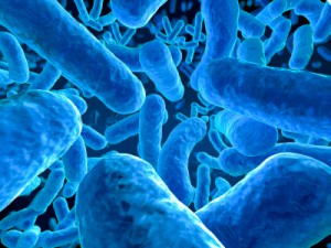
Every day scientists apply creative ideas to solve real-world problems. Every so often a paper comes up that highlights the creativity and elegance of this process in a powerful way. The paper “Programmable probiotics for detection of cancer in urine”, published May 27 in Science Translational Medicine, provides one great example of the application of scientific creativity to develop potential new ways for early detection of cancer.
The paper describes use of an engineered strain of E.coli to detect liver tumors in mice. The authors (Danino et al) developed a potential diagnostic assay that uses a simple oral delivery method and provides a readout from urine, all of which is made possible by some seriously complex and elegant science. Continue reading “Designer Bacteria Detect Cancer”
If you think back to biology class, you most likely learned that endothermy, or the ability to maintain a body favorable body temperature (i.e., different than the ambient temperature), was a unique characteristic of mammals and birds. This ability sets “warm blooded” animals apart from “cold blooded” ectotherms such as reptiles and fish.
For fish, one of the biggest challenges to maintaining an elevated body temperature is convective heat loss in the gill lamellae. There are a few fish species that are able to retain some of the heat generated internally, but these “regional endotherms” are only able to increase the temperature of specific areas or tissues. Regional endotherms limit the heat loss with retia mirabilia, which are a complex network of blood vessels that act as counter-current heat exchangers that warm the cold arterial blood as it returns from the gills. However, these retia have only been found associated with specific muscle groups and organs, and as a result the rest of the fish’s body remains at ambient temperature.
A silver and crimson-colored, tire-sized, fish called the opah (Lampris guttatus), or moonfish, is changing what scientist thought they knew about endothermy in fish (1). The opah lives in the cold, dim waters of the ocean’s mesopelagic zone hundreds of feet below the surface. Unlike other fish at these depths, the opah is a fast swimming, agile predator. Its secret is retia located inside the gills. The location of the retia means that the opah can maintain an elevated body temperature throughout its body, and a warmer body temperature means it can swim faster and react more quickly than both its prey and other predators.
I don’t think that we should throw out all our biology text books and start rewriting descriptions of what defines a fish, but the discovery of the warm blooded opah should remind us that what we know is just a small drop compared to what remains to be discovered.
Reference
Wenger, N. et al. (2015) Whole-body endothermy in a mesopelagic fish, the opah, Lampris guttatus. Science348, 786–9.
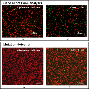
Figure 5 shows typical scanned images of bead-array for analyzing adjacent normal tissue and tumor tissue. Huang et al. (2015) Digital Detection of Multiple Minority Mutants and Expression Levels of Multiple Colorectal Cancer-Related Genes Using Digital-PCR Coupled with Bead-Array. PLOS ONE10(4):e0123420. doi:10.1371/journal.pone.0123420.g005The ideal cancer detection method would involve giving a sample of blood or tissue and using DNA or RNA analysis to determine if there were any gene sequence or gene expression changes that are known hallmarks of cancer. Unfortunately, most current screening methods used are not so precise and in some cases are invasive. However useful tests for colon cancer may be, many people do not subject themselves to the standard colonoscopy. What if there was an easier, noninvasive method that could be used to screen for cancer and detect changes at the early, easily treatable stages of cancer? A recent article in PLOS ONE describes just such a mutation detection method for colorectal cancer using purified nucleic acid with a method that involves emulsion PCR, bead arrays and fluorescent probes. Continue reading “Cancer Detection on a Chip?”
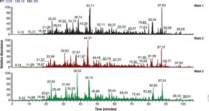
Filter-aided sample preparation (FASP) method is used for the on-filter digestion of proteins prior to mass-spectrometry-based analyses (1,2). FASP was designed for the removal of detergents, and chaotropes that were used for sample preparation. In addition, FASP removes components such as salts, nucleic acids and lipids. Akylation of reduced cysteine residues is also carried out on filter, after which protein is proteolyzed by use of trypsin on filter in the optimal buffer of the enzyme. Subsequent elution and desalting of the peptide-rich solution then provides a sample ready for LC–MS/MS analysis.
Erde et al. (3) described an enhanced FASP (eFASP) workflow that included 0.2% DCA in the exchange, alkylation, and digestion buffers,thus enhancing trypsin proteolysis, resulting in increases cytosolic and membrane protein representation. DCA has been reported (4) to improve the efficiency of the denaturation, solubilization, and tryptic digestion of proteins, particularly proteolytically resistant myoglobin and integral membrane proteins, thereby enhancing the efficiency of their identification with regard to the number of identified proteins and unique peptides.
In a recent publication (5) traditional FASP and eFASP were re-evaluated by ultra-high-performance liquid chromatography coupled to a quadrupole mass filter Orbitrap analyzer (Q Exactive). The results indicate that at the protein level, both methods extracted essentially the same number of hydrophobic transmembrane containing proteins as well as proteins associated with the cytoplasm or the cytoplasmic and outer membranes.
The LC–MS/MS results indicate that FASP and eFASP showed no significant differences at the protein level. However, because of the slight differences in selectivity at the physicochemical level of peptides, these methods can be seen to be somewhat complementary for analyses of complex peptide mixtures.
Pasque flowers in a northern hemisphere garden in spring.
As the seasons change so does the general state of health for many of us. The further from the equator we live, the more pronounced these effects are. For instance, did you know that blood pressure elevation for many people increases with the distance they live from the equator, an effect most pronounced during the low sunlight season (winter in the northern hemisphere)?
“Here we find more than 4,000 protein-coding mRNAs in white blood cells and adipose tissue to have seasonal expression profiles, with inverted patterns observed between Europe and Oceania.”
Let’s Take a Look at the Research
Todd et al. looked at ethnically and geographically distinct populations, including subjects from Australia, The Gambia (Africa), Germany, the UK and Iceland. Individuals from the various studies were infants, adults with type 1 diabetes and asthmatics in the range of 18-83 years of age. The authors analyzed RNA from peripheral blood mononuclear cells and subcutaneous adipose tissue biopsies, as well as examining peripheral blood cell counts and circulating levels of proinflammatory cytokines. Continue reading “Your Health has a Season”
There are times when I ask myself why I chose a career in science. This happens on what I call “grass is greener” days. On these days I dream of other careers—like National Geographic reporter or Caribbean tour guide–which all sound way more exciting than scientist. Admittedly these alternative careers are not ones that many people have the privilege of attaining, but sometimes reality gets to take a vacation. Fortunately, science is a fast-moving, always-changing field. As much as I might occasionally dream of exotic jobs in far away locations, science always pulls me back in with something new and unexpected. Because as much as we’d like to think we know, the truth is there is so much more that we don’t.
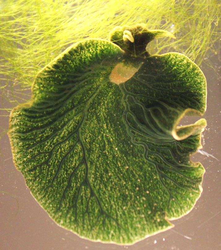
The sea slug Elysia chlorotica. Image from: Pelletreau K.N., et al. (2014) PLoS ONE 9: e97477.
A case in point—sea slugs. These unfortunately named, exotic looking creatures have some surprising secrets.
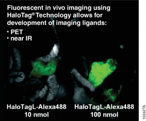
Antibodies labelled with radioisotopes or the sequential administrationof an antibody and a radioactive secondary agent facilitate the in vivo detection and/or characterisation of cancers by positron emission tomography (PET) or by single-photon emission computed tomography (SPECT) imaging.
There are drawbacks to both methods, including prolonged exposure to radiation and ensuring that both the antibody and the radiolabelled secondary agent are suitably designed so that they bind rapidly upon contact at the tumor.
A recent publication (1) investigated a alternative method utilizing the HaloTag® dehalogenase enzyme HaloTag® is a dehalogenase enzyme (33 kDa) that contains an engineered cavity designed to accommodate the reactive chloroalkane group of a HaloTag® ligand (HTL). Upon entering the enzyme cavity, the terminal chlorine atom rapidly undergoes nucleophilic displacement, and a covalent adduct is formed, effectively anchoring the HaloTag® ligand in a precise location.
Three new HaloTag® ligands were synthesized and each labelled with the SPECT radionuclide indium-111 111In-HTL-1 and the dual-modality HaloTag® ligands,111In-HTL-2 and111;In-HTL-3 containing TMR which allows complementary imaging data).
For the validation of the pretargeting strategy based on these HaloTag® ligands, the target human epidermal growth factor receptor 2 (HER2)was selected. Trastuzumab (Herceptin®) was selected as the primary targeting agent and was modified with HaloTag® protein via the trans-cyclooctene/tetrazine ligation.
All three 111In-labelled HaloTa®g ligands exhibited significantly higher binding to the HER2 expressing when compared to negative controls.
XWe use cookies and similar technologies to make our website work, run analytics, improve our website, and show you personalized content and advertising. Some of these cookies are essential for our website to work. For others, we won’t set them unless you accept them. To learn more about our approach to Privacy we invite you to Read More
By clicking “Accept All”, you consent to the use of ALL the cookies. However you may visit Cookie Settings to provide a controlled consent.
We use cookies and similar technologies to make our website work, run analytics, improve our website, and show you personalized content and advertising. Some of these cookies are essential for our website to work. For others, we won’t set them unless you accept them. To find out more about cookies and how to manage cookies, read our Cookie Policy.
If you are located in the EEA, the United Kingdom, or Switzerland, you can change your settings at any time by clicking Manage Cookie Consent in the footer of our website.
Necessary cookies are absolutely essential for the website to function properly. These cookies ensure basic functionalities and security features of the website, anonymously.
Cookie
Duration
Description
cookielawinfo-checbox-analytics
11 months
This cookie is set by GDPR Cookie Consent plugin. The cookie is used to store the user consent for the cookies in the category "Analytics".
cookielawinfo-checbox-functional
11 months
The cookie is set by GDPR cookie consent to record the user consent for the cookies in the category "Functional".
cookielawinfo-checbox-others
11 months
This cookie is set by GDPR Cookie Consent plugin. The cookie is used to store the user consent for the cookies in the category "Other.
cookielawinfo-checkbox-advertisement
1 year
The cookie is set by GDPR cookie consent to record the user consent for the cookies in the category "Advertisement".
cookielawinfo-checkbox-necessary
11 months
This cookie is set by GDPR Cookie Consent plugin. The cookies is used to store the user consent for the cookies in the category "Necessary".
cookielawinfo-checkbox-performance
11 months
This cookie is set by GDPR Cookie Consent plugin. The cookie is used to store the user consent for the cookies in the category "Performance".
gdpr_status
6 months 2 days
This cookie is set by the provider Media.net. This cookie is used to check the status whether the user has accepted the cookie consent box. It also helps in not showing the cookie consent box upon re-entry to the website.
lang
This cookie is used to store the language preferences of a user to serve up content in that stored language the next time user visit the website.
viewed_cookie_policy
11 months
The cookie is set by the GDPR Cookie Consent plugin and is used to store whether or not user has consented to the use of cookies. It does not store any personal data.
Analytical cookies are used to understand how visitors interact with the website. These cookies help provide information on metrics the number of visitors, bounce rate, traffic source, etc.
Cookie
Duration
Description
SC_ANALYTICS_GLOBAL_COOKIE
10 years
This cookie is associated with Sitecore content and personalization. This cookie is used to identify the repeat visit from a single user. Sitecore will send a persistent session cookie to the web client.
vuid
2 years
This domain of this cookie is owned by Vimeo. This cookie is used by vimeo to collect tracking information. It sets a unique ID to embed videos to the website.
WMF-Last-Access
1 month 18 hours 24 minutes
This cookie is used to calculate unique devices accessing the website.
_ga
2 years
This cookie is installed by Google Analytics. The cookie is used to calculate visitor, session, campaign data and keep track of site usage for the site's analytics report. The cookies store information anonymously and assign a randomly generated number to identify unique visitors.
_gid
1 day
This cookie is installed by Google Analytics. The cookie is used to store information of how visitors use a website and helps in creating an analytics report of how the website is doing. The data collected including the number visitors, the source where they have come from, and the pages visted in an anonymous form.
Advertisement cookies are used to provide visitors with relevant ads and marketing campaigns. These cookies track visitors across websites and collect information to provide customized ads.
Cookie
Duration
Description
IDE
1 year 24 days
Used by Google DoubleClick and stores information about how the user uses the website and any other advertisement before visiting the website. This is used to present users with ads that are relevant to them according to the user profile.
test_cookie
15 minutes
This cookie is set by doubleclick.net. The purpose of the cookie is to determine if the user's browser supports cookies.
VISITOR_INFO1_LIVE
5 months 27 days
This cookie is set by Youtube. Used to track the information of the embedded YouTube videos on a website.
Performance cookies are used to understand and analyze the key performance indexes of the website which helps in delivering a better user experience for the visitors.
Cookie
Duration
Description
YSC
session
This cookies is set by Youtube and is used to track the views of embedded videos.
_gat_UA-62336821-1
1 minute
This is a pattern type cookie set by Google Analytics, where the pattern element on the name contains the unique identity number of the account or website it relates to. It appears to be a variation of the _gat cookie which is used to limit the amount of data recorded by Google on high traffic volume websites.

 Every day scientists apply creative ideas to solve real-world problems. Every so often a paper comes up that highlights the creativity and elegance of this process in a powerful way. The paper “Programmable probiotics for detection of cancer in urine”, published May 27 in Science Translational Medicine, provides one great example of the application of scientific creativity to develop potential new ways for early detection of cancer.
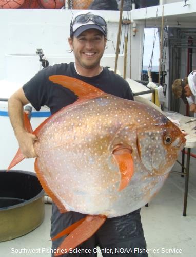
 Filter-aided sample preparation (FASP) method is used for the on-filter digestion of proteins prior to mass-spectrometry-based analyses (1,2). FASP was designed for the removal of detergents, and chaotropes that were used for sample preparation. In addition, FASP removes components such as salts, nucleic acids and lipids. Akylation of reduced cysteine residues is also carried out on filter, after which protein is proteolyzed by use of trypsin on filter in the optimal buffer of the enzyme. Subsequent elution and desalting of the peptide-rich solution then provides a sample ready for LC–MS/MS analysis.
 Antibodies labelled with radioisotopes or the sequential administrationof an antibody and a radioactive secondary agent facilitate the in vivo detection and/or characterisation of cancers by positron emission tomography (PET) or by single-photon emission computed tomography (SPECT) imaging.