Beating the Odds of Cancer: Not Just a Tall Tale

elephants_web
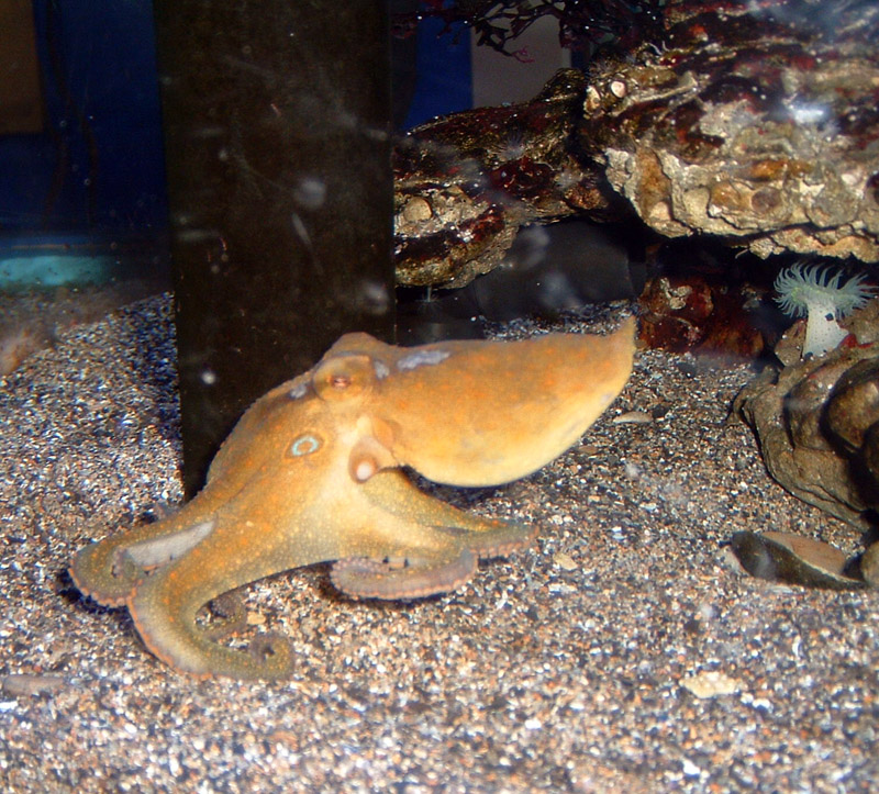
When it comes to combating cancer does size matter? If every cell in the body has the propensity to become cancerous, it should naturally follow that larger animals that pack greater number of body cells and that those whose cells undergo greater number of cell divisions are more likely to develop cancer. By the same logic, organisms with longer lifespans must also have a greater chance of accumulating mutations leading to cancer. Surprisingly, the risk of developing cancer is only 5% in elephants and 18% in whales whereas it is as high as 30% in humans and rodents.  The apparent lack of correlation between body mass, longevity and cancer- known as Peto’s paradox- has flummoxed scientists for several decades.1

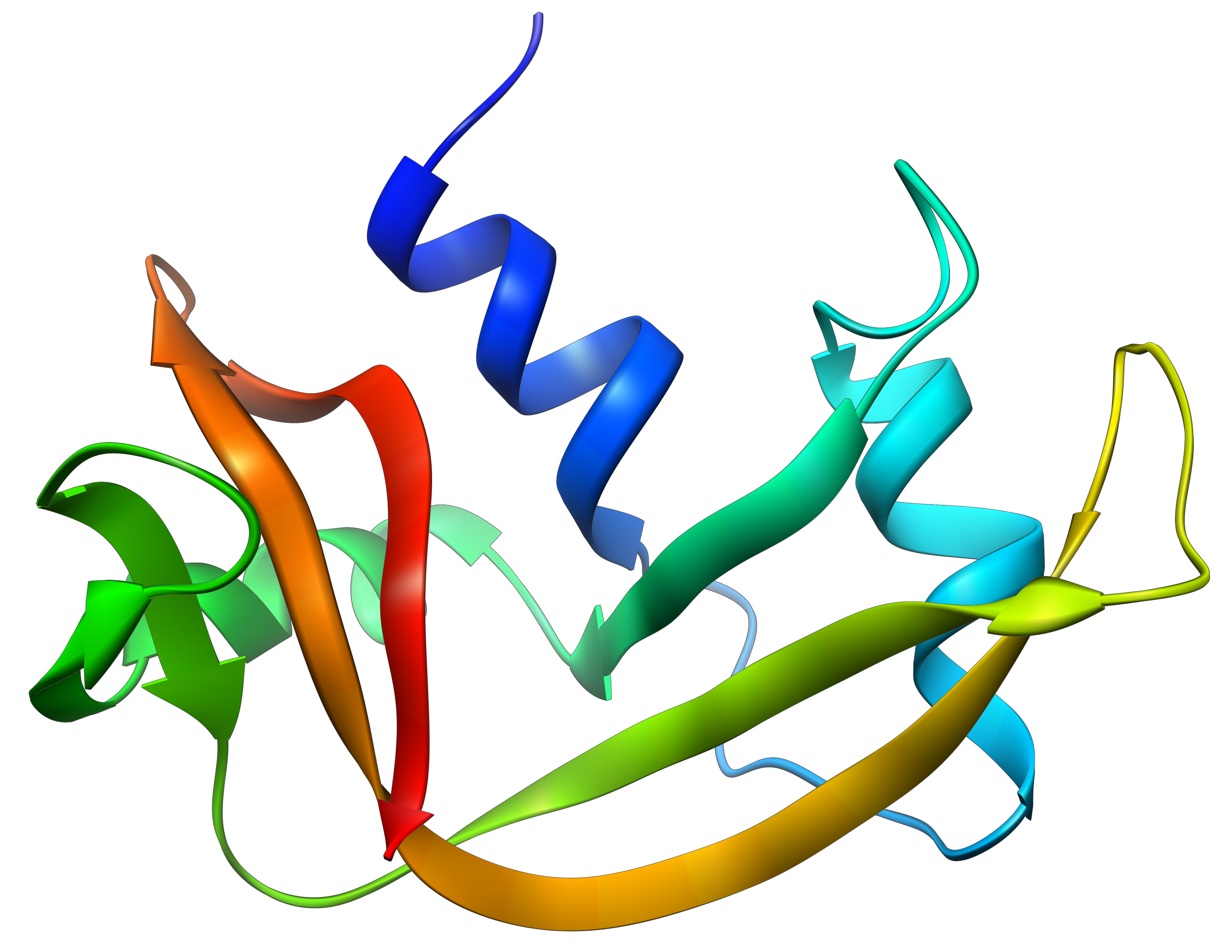
A recent study published in Journal of the American Medical Association  by  Abegglen and colleagues has unlocked the secret weapon held by the pachyderms in fighting cancer2. While the weapon itself might not be new to cancer biologists, the stash carried by these marvelous animals is the highest recorded for any living species so far. To understand this weapon let’s revisit the coping mechanisms developed by cells to prevent cancer. When mammalian cells are exposed to cancer inducing treatments, such as UV radiation for example, a gene encoding TP53, kicks into gear making copies of the tumor suppressing protein of the same name. TP53 acts as a tumor suppressor, which means that it regulates cell division by keeping cells from growing and dividing too fast or in an uncontrolled way. It does so by either repairing any damage to the cells caused by the UV exposure or by killing off the cell by a self-destructing mechanism known as apoptosis which is akin to committing suicide.

Many mammals, including humans carry only two copies of this important gene; one copy or allele is inherited from each parent. If the TP53 gene is inactivated by mutations, the risk of developing cancer increases by several fold. A rare but lethal condition called Li-Fraumeni Syndrome marks patients who have only one working copy of TP53 with more than a 90 percent lifetime cancer risk from childhood into their adult years.  In a quest to investigate the unexplained resistance to cancer by elephants, the scientists combed through the elephant genome and stumbled upon 40 copies of genes that code for TP53. One pair was ancestral in origin, whereas the remainder appear to have diverged from the ancestral copy and were archived within the genome over the course of evolution as retrogenes. Continue reading “Beating the Odds of Cancer: Not Just a Tall Tale”

Noninvasive Prenatal Genetic Testing Using Circulating Cell-Free DNA

Expectant MotherScientists have known for some time that fetal DNA can be detected in the maternal bloodstream during pregnancy (1). Up to 10% of circulating cell-free-DNA (ccfDNA) can be attributed to the fetus. Fetal ccfDNA is released from the placenta, mainly through apoptosis, and enters the maternal bloodstream, where it can be easily collected and detected by PCR amplification. This method of collection has a much lower risk of miscarriage compared to more invasive collection methods such as amniocentesis and chorionic villus sampling.

Amplification of fetal ccfDNA enables a number of prenatal genetic testing such as gender determination and detection of fetal aneuploidy and other mutations. Testing of ccfDNA also allows identification of fetuses with a higher risks of hemolytic disease of the newborn (erythroblastosis fetalis) due to expression of the Rh factor in an Rh– negative mother, who can develop antibodies against the Rh factor and mount an immune response against fetal red blood cells. Finally, ccfDNA allows prenatal paternity determination (2). However, these tests have limitations.

Continue reading “Noninvasive Prenatal Genetic Testing Using Circulating Cell-Free DNA”

A Potential Single-Tube Multiplex Assay for Detecting Dengue Virus in the Field

In areas of the world where the electricity is intermittent, resources are limited and transporting bulky equipment and reagents that are sensitive to temperature fluctuations is difficult, diagnosis of viruses like dengue can be challenging. If you could reduce or eliminate the need for electricity dependent equipment for diagnostic assays without sacrificing sensitivity or specificity, it would be a boon to field workers. An article published in PLOS ONE describes how researchers developed a multiplex isothermal amplification method that could assess a potential dengue infection with a visual real-time or endpoint detection in a single tube.

Countries affected by dengue. By Percherie (Distribution de la dengue sur Commons) [GFDL (http://www.gnu.org/copyleft/fdl.html), CC-BY-SA-3.0 (http://creativecommons.org/licenses/by-sa/3.0/) or CC BY-SA 2.5-2.0-1.0 , via Wikimedia Commons
Continue reading “A Potential Single-Tube Multiplex Assay for Detecting Dengue Virus in the Field”

Rewriting the Histone Code: Searching for Treatments for Stage IV Thyroid Cancers

Chromatin fiberOften a diagnosis of thyroid cancer is associated with a good prognosis and fairly straightforward surgical treatments to remove the tumor followed by radioactive iodine ablation. Such treatment works well in tumors that have not metastasized and retain enough of their thyroid cell “identity” that they can still accumulate radioactive iodine.

However, aggressive thyroid cancers, which often metastasize and recur, do not respond to standard treatments because they are generally too dedifferentiated to accumulate iodine, so alternative treatments are needed.

One approach is to look for compounds that will reverse dedifferentiation, making tumor cells more likely to take up and concentrate radioactive iodine regardless of their location in the body. One possible target to effect dedifferentiation is epigenetic modification of histone proteins.

Histone proteins are more than the structural components of the nucleosome that organizes the chromatin inside cells. Histone proteins are subject to a host of protein modifications on their N-terminal tails such as acetylation, phosphorylation, methylation, ubiquitination and ADP-ribosylation. These various modifications are seen as creating a “histone code” that is read by other proteins and protein complexes (1). This code regulates patterns of gene expression and activity for a cell—in part resulting in a differentiated phenotype. Previous studies have suggested that some histone deacetylase (HDAC) inhibitors (e.g., valproic acid) can reverse some of the dedifferentiation associated with aggressive cancers (2).

Jang, et al. in a recent paper (3) published in Cancer Gene Therapy synthesized a group of HDAC inhibitor analogs (AB1–AB13) and tested them for their ability to inhibit growth of three aggressive human thyroid cancer cell lines and induce partial re-differentiation to the thyroid cell phenotype. Continue reading “Rewriting the Histone Code: Searching for Treatments for Stage IV Thyroid Cancers”

A Normalization Method for Luciferase Reporter Assays of miRNA-Mediated Regulation

Today’s blog is from guest blogger Ken Doyle of Loquent, LLC. Here, Ken reviews a 2014 paper highlighting specific considerations for using reporter assays to study miRNA-mediated gene regulation.

mirna

The accelerated pace of research into noncoding RNAs has revealed multiple regulatory roles for microRNAs (miRNAs). These diminutive noncoding RNA species—typically 20-24 nucleotides in length—are now known to mediate a broad range of biological functions in plants and animals. In humans, miRNAs have been implicated in various aspects of development, differentiation, and metabolism. They are known to regulate an assortment of genes involved in processes from neuronal development to stem cell division. Dysregulation of miRNA expression is associated with many disease states, including neurodegenerative disorders, cardiovascular disease, and cancer.

Typically, miRNAs act as post-transcriptional repressors of gene expression, either by targeted degradation of messenger RNA (mRNA) or by interfering with mRNA translation. Most miRNAs exert these effects by binding to specific sequences called microRNA response elements (MREs). These sequences are found most often within the 3´-untranslated regions (3´-UTRs) of animal genes, while they may occur within coding sequences in plant genes.

Studies of the regulatory roles played by miRNAs often involve cell-based assays that use a reporter gene system, such as luciferase or green fluorescent protein. In a standard assay, the reporter gene is cloned upstream of the 3´-UTR sequence being studied; this construct is then cotransfected with the miRNA into cells in culture. A study by Campos-Melo et al., published in September 2014, examined this experimental approach for miRNAs from spinal cord tissues, using firefly luciferase as the reporter gene and Renilla luciferase as a transfection control.

Continue reading “A Normalization Method for Luciferase Reporter Assays of miRNA-Mediated Regulation”

Sequencing the Octopus Genome: Invertebrate Intelligence Explained?

In a Letter in Nature magazine last week (August 13, 2015), researchers published surprising findings from a genome analysis of the octopus. As a result, we now know that this invertebrate has more than just behavioral oddities with which to amaze.

In their publication, C. Albertin et al. report the results of genome sequencing of the California two-spot octopus, Octopus bimaculoides. They did not find the predicted whole-genome duplication, but rather an unexpectedly large genome with many rearrangements, and two gene family expansions that were previously thought to exist only in vertebrates.

Califonia two spot octopus. Image by Jeremy S. Taken at Santa Monica Aquarium. Used via Creative Commons license, Wikimedia.

The Research
Albertin et al. sequenced the O. bimaculoides genome using a whole-genome shotgun approach, and then annotated it using extensive transcriptome sequences from 12 tissues. They estimate that the genome assembly incorporated 97% of protein-coding sequences, and 83% of the entire 2.7gigabase genome. The remaining sequence was composed largely of repetitive elements. Continue reading “Sequencing the Octopus Genome: Invertebrate Intelligence Explained?”

Manipulating Microbiota: A Synthetic Biology Exploration of the Gut

33553646_l

Microbial cells outnumber the cells of our own bodies approximately 10:1, these microbes that live on our skin and along the epithelial linings of our internal tubes make up our microbiota*, and they can have major effects on our health. Most of our microbiota are commensal organisms, living in harmony with our body, but if you suppress our immune system or greatly reduce their populations with large doses of antibiotics, and you will soon see the effects of disrupting our microbiota.

There is much interest in the microbiota that inhabit our bodies. For instance several studies have indicated that intestinal microbes can play a big part in obesity, with changes in the makeup of the microbiota being a major risk factor (1). But many of these organisms are hard to learn about—the ones that inhabit the deep folds of our gut thrive in moist, warm, anaerobic conditions with lots of specialized nutrients, conditions that are very hard to replicate in the laboratory. For that reason, we don’t know much about many of the microbes that are the most abundant within us.

The Human Microbiome Project begun in 2008 by the National Institutes of Health (2) seeks to understand human microbiota and their relationship to human health. To do this, the researchers leading the project took a metagenomic approach—using advanced DNA sequencing technologies to sequence the genomes of human microbiota and get a look at the human microbiome—without culturing the microbes.

But to truly understand their biology, and to perhaps exploit what we learn to enhance human health we need to be able to manipulate these organisms. In particular, biologists who are interested in synthetic biology would like to use these micro-organisms to monitor what is going on in our bodies, particularly our guts. What better monitor for these hard-to-access places than an organism that is already well adapted to live there? 

Continue reading “Manipulating Microbiota: A Synthetic Biology Exploration of the Gut”

Molecular Autopsies in the Whole Genome Sequencing Era

Engraving of the human heart by T. Milton, 1814. Image courtesy of Wikimedia Commons. In some cases of death, molecular autopsies are the best way to understand cause of death.
Engraving of the human heart by T. Milton, 1814. Image courtesy of Wikimedia Commons.

Every year, nearly 8 million people die from sudden cardiac death, which is defined as the unexpected death of a seemingly healthy person due to malfunctions in the heart’s electrical system and loss of cardiac function. Although sudden cardiac death (SCD) is usually associated with mature adults, SCD claims thousands of young lives every year. In most cases, the cause of death can be determined by autopsy or toxicological analysis, but up to 30% of these premature deaths have no apparent cause, leaving medical examiners and family members of the young victims to wonder what happened.

In cases where traditional pathological examinations cannot provide insight into causation, medical examiners are increasingly turning to molecular autopsies to determine if there is an underlying genetic factor that contributed to a person’s death.

Continue reading “Molecular Autopsies in the Whole Genome Sequencing Era”

All You Need is Pla (for Pneumonic Plague)

Yersinia pestis. By Mrs Robinson at bg.wikipedia (Transferred from bg.wikipedia) [Public domain], from Wikimedia Commons.
Writing about Yersinia pestis or the Black Death, has earned me a reputation among Promega Connections bloggers. I am interested in what researchers have been able to piece together about the causative agent of ancient plagues, what modern research shows about how Y. pestis spreads in the body and the continuing reservoirs in modern times, resulting in publication of eight blog posts on the subject. Understanding Y. pestis bacterium is of continuing interest to researchers. How did Yersinia pestis evolve from the humble Yersinia pseudotuberculosis, a pathogen that causes gastrointestinal distress, into a virulent pneumonic plague that is a global killer? One strategy for answering this question is to look at the genomic tree of Y. pestis and trace which strains had what characteristics. In a recent Nature Communications article, Zimbler et al. explored the role of the plasmid pPCP1 in Y. pestis evolution and the signature protease Pla it expresses. Continue reading “All You Need is Pla (for Pneumonic Plague)”

A Reason for Ribonuclease: From Laboratory Nuisance to Cancer Therapeutic

"RNase A". Licensed under CC BY-SA 2.5 via Wikimedia Commons - https://commons.wikimedia.org/wiki/File:RNase_A.png#/media/File:RNase_A.png
“RNase A”. Licensed under CC BY-SA 2.5 via Wikimedia Commons – https://commons.wikimedia.org/wiki/File:RNase_A.png#/media/File:RNase_A.png

RNase, back in the early 1990s, posed a serious threat to laboratories working with RNA isolation. My graduate work involved isolating RNA from the tissues of Lyme disease-infected mice and hamsters. We struggled to DEPC-treat glass and plasticware, or autoclave anything that could be autoclaved, kept tissues cold during RNA harvest and held our breaths (truly, as aerosol could be another source of ribonuclease) until PAGE proved us successful in RNA isolation.

Ribonuclease (RNase) was omnipresent and the arch rival of our work, across several species, due to its RNA destroying abilities.

Now, a July 13, 2015 publication by researchers at the University of Wisconsin-Madison provided both a catch-up for this former lab rat on modern day research with and knowledge of RNase, as well as an exciting look at what may be a real purpose for this RNA-destroying molecule: RNase has moved to clinical trials due to the discovery of it’s cytotoxicity for cancer cells.

Raines’ group in the Department of Chemistry at UWI-Madison published in ACS Central Science their findings on the ligand that RNase 1 uses to attach to human cancer cells, in the article, “Human Cancer Antigen Globo H is a Cell-Surface Ligand for Human Ribonuclease 1”. Continue reading “A Reason for Ribonuclease: From Laboratory Nuisance to Cancer Therapeutic”