Finding a Connection Between Glucose Metabolism and Macrophage Activation

Introduction to Glucose Metabolism

Macrophages. By NIAID (https://www.flickr.com/photos/niaid/17380707492/) [CC BY 2.0 (http://creativecommons.org/licenses/by/2.0)], via Wikimedia Commons
Many think of glucose as something diabetics have to test each day using a blood monitor, or a quick energy boost for someone exercising intensely. However, the simple sugar glucose, a monosaccharide, fuels most of the cells in our bodies. Disaccharides that contain glucose (e.g., sucrose is comprised of glucose and fructose) and glucose polymers (e.g., starch and glycogen) are carbohydrates that are consumed by organisms from bacteria to humans to produce energy. These carbohydrates are broken down into component monosaccharides like glucose and lactose. The process of glycolysis generates the energy currency of cells, ATP, as well as precursor molecules for nucleotides, lipids and amino acids. Because glucose is the cell fuel source, the uptake of glucose and its subsequent metabolism is increased by cells that divide rapidly like cancer cells. The more energy and precursor molecules the cancer cell can create for itself, the more rapidly the tumor can grow.

Because glucose metabolism is central to cellular functioning, changes that decrease glucose uptake or increase glycolysis have a widespread effect on on both the cells and organism. How does a simple sugar molecule create such broad effects on health? For example, diabetes results from the inability to store glucose because of a lack of insulin, a hormone that draws glucose from the blood and stores it as glycogen in the liver, muscles and adipose tissue. High levels of sugar in the blood negatively affect the body over the long term, damaging blood vessels and eyesight, making the kidneys work harder to excrete the excess sugar and increasing the risk of stroke and coronary artery disease. Because cancer cells have such a high metabolic demand for glucose, many of the mutations in cancers affect pathways that regulate glucose uptake and glucose breakdown, allowing the cancer cells to survive and grow, crowding out nearby normal cells.

Glucose metabolism is altered by processes other than mutations or an reduced production of a hormone. Throughout its life cycle, a cell will vary its requirements for glucose. For example, the cells that comprise our innate immune response are typically in a quiescent or steady state. However, when these immune cells encounter an foreign invader, they become activated and increase their demand for glucose. To respond to a potential pathogen, the activated cells need glucose to fuel cell proliferation and the production of cytokines, chemicals that activate other immune cells and initiate an inflammatory response. The typical signs of inflammation are red inflamed area that may be painful to the touch, such as a cut that becomes infected. Most inflammation resolves when the infection is eliminated, leaving behind whole skin in the instance of a cut, and the activated immune cells become quiescent again.

An Interesting Observation about Glucose Metabolism in M2 Macrophages

Glucose uptake, immunity and metabolism are cellular pathways that are intertwined such that understanding how glucose is utilized in macrophages illuminates gene induction and regulation in activated macrophages. In a recently published eLife article, Covarrubias et al. studied how activation of murine bone marrow-derived macrophages (BMDMs) by interleukin-4 (IL-4), a signaling cytokine, altered glucose metabolism in the cells and regulated a subset of genes involved in macrophage activation. Continue reading “Finding a Connection Between Glucose Metabolism and Macrophage Activation”

Vitamin D: Power in Cancer Prevention?

This and vitamin D should get your attention.

Have you ever noticed that after a good long day outdoors, maybe hiking, at the beach or even working in the yard, you feel really strong and healthy, maybe even more relaxed than after an indoor session in front of the telly or computer? Maybe a February trip to someplace sunny like Mexico or the Canary Islands has given you renewed zest for your normal tasks?

While rest and a change of scenery is never a bad thing, time outdoors and in the sunshine might have gained for you something more than rest and relaxation. If it included a little UVB irradiation, your time outdoors may have increased your serum vitamin D level. And though it’s been presumed for years, we now have proof that higher serum vitamin D3 levels correlate with a decreased incidence of certain cancers. Continue reading “Vitamin D: Power in Cancer Prevention?”

Increasing Drug Research and Development Efficiency Using a 4-point Screening Method to Determine Molecular Mechanism of Action

Fig 4. Four point MMOA screen for tideglusib and GW8510. Time dependent inhibition was evaluated by preincubation of TbGSK3β with 60 nM tideglusib and 6 nM GW-8510 with 10μM and 100μM ATP. (A). Tideglusib [60 nM] in 10μM ATP. (B). GW8510 [60 nM] in 10μM ATP. (C.) Tideglusib [60 nM] at 100μM ATP. (D.) GW8510 [60 nM] at 100μM ATP. All reactions preincubated or not preincubated with TbGSK3β for 30 min at room temperature. Experiments run with 10μM GSM peptide, 10μM ATP, and buffer. Minute preincubation (30 min) was preincubated with inhibitor, TbGSK3β, GSM peptide, and buffer. ATP was mixed to initiate reaction. No preincubation contained inhibitor, GSM peptide, ATP, and buffer. The reaction was initiated with TbGSK3β. Reactions were run at room temperature for 5 min and stopped at 80°C. ADP formed was measured by ADP-Glo kit. Values are mean +/- standard error. N = 3 for each experiment and experiments were run in duplicates. Control reactions contained DMSO and background was determined using a zero time incubation and subtracted from all reactions. Black = 30 min preincubation Grey = No preincubation.
Four point MMOA screen for tideglusib and GW8510.
Time dependent inhibition was evaluated by preincubation of TbGSK3β with 60 nM tideglusib and 6 nM GW-8510 with 10μM and 100μM ATP. (A). Tideglusib [60 nM] in 10μM ATP. (B). GW8510 [60 nM] in 10μM ATP. (C.) Tideglusib [60 nM] at 100μM ATP. (D.) GW8510 [60 nM] at 100μM ATP. All reactions preincubated or not preincubated with TbGSK3β for 30 min at room temperature.  Black = 30 min preincubation Grey = No preincubation.
The first small-molecule kinase inhibitor approved as a cancer therapeutic, imatinib mesylate (Gleevec® treatment), has been amazingly successful. However, a thorough understanding of its molecular mechanism of action (MMOA) was not truly obtained until more than ten years after the molecule had been identified.

Understanding the MMOA for a small-molecule inhibitor can play a major role in optimizing a drug’s development. The way a drug actually works–the kinetics of binding to the target molecule and how it competes with endogenous substrates of that target–ultimately determines whether or not a a candidate therapeutic can be useful in the clinic. Drugs that fail late in development are extremely costly.

Drug research and discovery for neglected tropical diseases suffer from a lack of a large commercial market to absorb the costs of late-stage drug development failures. It becomes very important to know as much as possible, simply and quickly, about MMOA for candidate molecules for these diseases that are devastating to large populations.

One such neglected topical disease is Human African trypanosomiasis (HAT, also known as sleeping sickness). Continue reading “Increasing Drug Research and Development Efficiency Using a 4-point Screening Method to Determine Molecular Mechanism of Action”

Analyzing the Effects of Yersinia pestis Infection on Gene Expression

Yersinia pestis. See page for author [Public domain], via Wikimedia Commons
While scientists using ancient DNA analysis are learning how Yersinia pestis developed over time into the causative agent of three worldwide pandemics, there is still much to learn about the early hours and days of an organism infected with the plague. Y. pestis still infects humans so any insight into disease progression is useful for determining treatment timing and even developing novel treatments to supplement or replace antibiotics. A 2012 study observed how Y. pestis injected into mice spread throughout the body using bioluminescent imaging to track the infection. More recent research reported in PLOS ONE used a more real-world route of infection by introducing an aerosolized Y. pestis to a nonhuman primate model and tracking the transcripts altered during the first 42 hours of infection. Continue reading “Analyzing the Effects of Yersinia pestis Infection on Gene Expression”

Optimizing Antibody Internalization Assays: pHAb Dyes

22255190_pHabAmine_3D_image_050615-final-large

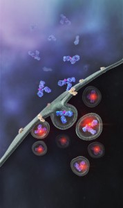
Promega has recently developed a method that allows antibodies to be screened for their internalization properties in a simple, plate-based format. The method uses pH sensor dyes (pHAb dyes), which are not fluorescent at neutral pH but become highly fluorescent at acidic pH. When an antibody conjugated with pHAb dye binds to its antigen on the cancer cell membrane, the antibody-dye-antigen complex is not fluorescent, but upon internalization and trafficking into endosomal and lysosomal vesicles the pH drops, and the dye becomes fluorescent.

To demonstrate the broad utility of the pHAb dye for receptor mediated antibody internalization, two therapeutic antibodies, trastuzumab and cetuximab,which bind to HER2 and EGFR respectively, were selected for a case study (1). Both the antibodies, which are known to internalize were labeled with pHAb dyes using amine or thiol chemistry.

Parameters such as the impact of dye–to-antibody ratio on the antigen–antibody binding, change in fluorescence as a function of pH of free dye and labeled dye, and labeled antibody internalization as a function of pHAb conjugated antibody concentration were evaluated.

The results indicate that pHAb dyes are pH sensitive fluorescent dyes that enable the study of receptor-mediated antibody internalization.Internalization assays can be performed in a plate-based homogeneous format and allow endpoint assays as well as real-time monitoring of internalization. They further show that internalization can be monitored even at a very low amount of antibody which is very important during the early monoclonal antibody development phase when the amount of sample is limited and the antibody concentration in the samples is low. a complimentary approach, they  also showed that a secondary antibody labeled with pHAb dye can be used instead of labeling primary antibodies.

Literature cited

Nath, N. et al. (2016) Homogeneous plate based antibody internalization assay using pH sensor fluorescent dye J.  Immunol. Methods epub ahead of print

Making Drug Discovery More Efficient: Predicting Drug Side Effects in Early Screening Efforts

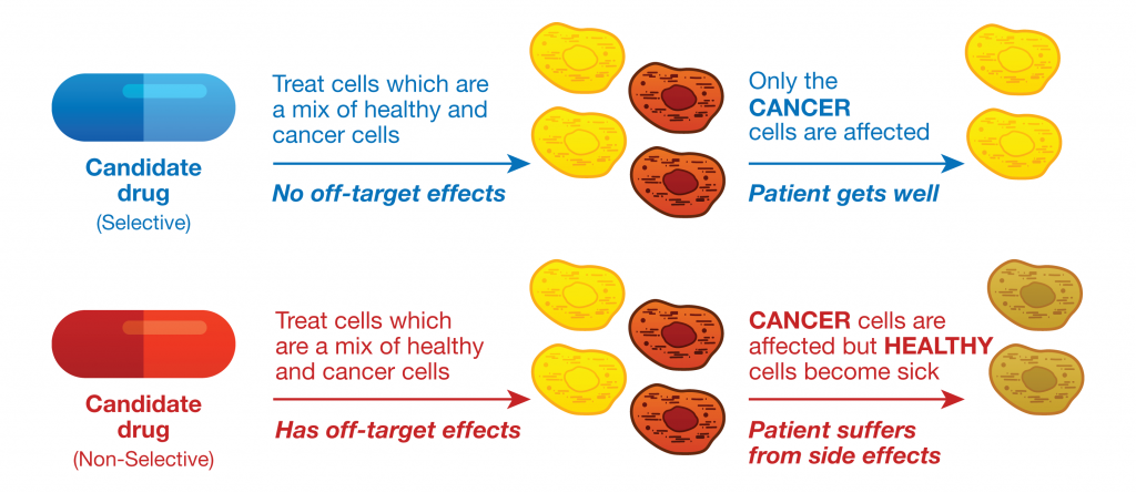
26911030-Laymans-KSPS-figure-WEB-R4Drug research and development is a complex and expensive process that begins with initial screening steps of candidate chemical compounds, and compounds that appear to have the desired potency against a specific cellular target or pathway are further evaluated. Candidate compounds that fail late in development or during clinical trials because of off-target effects are costly, and can be dangerous. Therefore drug developers not only need to ensure that a candidate compound is effective as a therapy, but also they need to predict any potential undesirable side effects due to off-target activities as early as possible in the drug discovery and development process. Continue reading “Making Drug Discovery More Efficient: Predicting Drug Side Effects in Early Screening Efforts”

To Meditate Perchance to Dream

First the disclosure: this blog is of course about Me.

But it’s also about You. And yours. Because as you know, we’ve become a culture that does not sleep.

Why don’t we sleep? I like to think that it is an evolutionary adaptation; not sleeping, after all, allows us more time for Facebook.

Or Etsy for you makers. Or Amazon for you shoppers. And let’s not forget our middle, high school and college students. Do they even have classrooms anymore, or are lectures all online (on screens)?

One tired pony. By Rachel C from Scotland (Flickr) [CC BY 2.0 (http://creativecommons.org/licenses/by/2.0)], via Wikimedia Commons
Honestly, the evolutionary adaptation idea comes from how we live and work today. And no, this is not another rant/lecture on the color of light emitted by whatever non-cathode ray tubes are in our phones or tablet-like devices.

It’s just that just working in our very busy online/wired world, jumping from web page to project management software, to big-screens in meetings has us adapted to being  on: capital “O” capital “N”.

This multi-multitasking has grown (for me) a new type of neurons that are not happy unless they are gleaning new information from a screen, all the time. And these neurons don’t stop working when the screen is gone; no, they continue seeking and trying to process. For me, if there’s no screen to look at, the neurons ping-pong around behind my eyeballs, looking and searching, as if to say, “Input missing! Input missing!”

The result can be hours in bed sans sleep; it seems the racket these neurons make keeps all the other neurons up. Continue reading “To Meditate Perchance to Dream”

What’s for Dinner? Mystery Meat Served at the 47th Explorer’s Club Annual Dinner Finally Identified

Could this be the 1951 mystery meat?

I remember one particular encounter with “mystery meat” when I was in college. I was walking along the serving line at the dining hall, and when I came to the entrée, I asked the server, “What is it?”

She replied quite succinctly, “Don’t know. Got beef in it.” I passed on the entrée that night, settling for salad and bread.

I would probably not have be a good candidate for membership in the Explorers Club.

The Explorers Club, founded in New York City in 1904, is a professional society that champions the cause of field research (1). The member list is impressive, including Teddy Roosevelt, the American President responsible for setting aside many of the most treasured public lands in the United States so that explorers have fields for research and wild places for adventures, Neil Armstrong, the first man to set foot on the moon, and Don Walsh and Jacques Piccard, the two men who descended into the Mariana’s trench to explore the deepest part of the ocean, among others.

In addition to a membership list that reads like a who’s who of science and exploration, The Explorers Club also has an annual dinner that for many years has popularized a menu of “exotic” foods (at least exotic foods from the point of view of the typical Midwest United States pallet). One of the club’s most celebrated dinners took place on January 13, 1951.

Continue reading “What’s for Dinner? Mystery Meat Served at the 47th Explorer’s Club Annual Dinner Finally Identified”

Inflammasomes: Peeking Inside the Inflammatory Process

Most of us have experienced an inflammatory response at some point in our lives. Fever, achy joints, swelling around a scrape or cut, all of these are forms of inflammatory response. Inflammation is the body’s response to infection or tissue damage and acts to limit harm to the rest of the body. A key player in the inflammation process is a group of protein complexes call inflammasomes. The term “inflammasome” was first used in 2002 by researchers in Switzerland (1) to refer to a caspase-activating protein complex. We now know that inflammasomes are cytosolic multiprotein platforms that assemble in response to pathogens and other signals. Inflammasome assembly results in the processing of the inactive procaspase-1 into the active cysteine-protease enzyme, caspase-1. Caspase-1, in turn, activates the proinflammatory cytokines Interleukins IL-1β and IL-18. In addition, caspase-1 is also required for pyroptosis, which is an inflammatory form of cell death that combines the characteristics of apoptosis (fragmented DNA) and necrosis (inflammation and cytokine release) and is frequently associated with microbial infections.

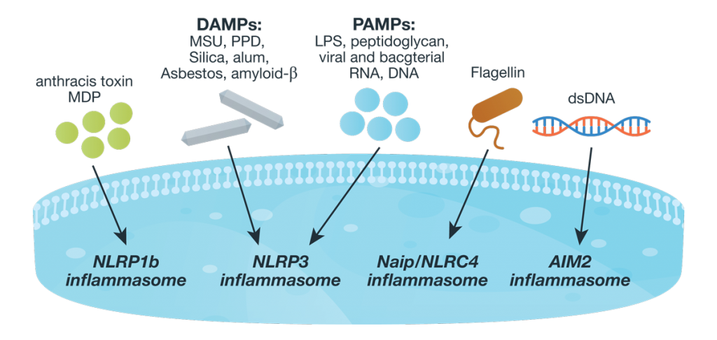
Inflammasome complexes are made up of scaffolding sensor proteins (NLR, AIM2, ALR), and an adaptor protein containing a caspase activation and retention domain (CARD) and inactive procaspase-1. Most inflammasomes are formed with one or two NLRs (NOD-like receptors). However, non-NLR proteins such as AIM2 (absent in melanoma 2) and pyrin can also form inflammasomes. The different sensor proteins are activated by different types of outside stimuli, and inflammasomes are loosely sorted into families based on the protein forming these sensors.26061534-Inflamasome-Assay-Blog-figure-WEB Continue reading “Inflammasomes: Peeking Inside the Inflammatory Process”

The Black Death: World Traveler or Persistent Homebody?

Spread of the Black Death. By Timemaps (Own work) [CC BY-SA 3.0 (http://creativecommons.org/licenses/by-sa/3.0) or GFDL (http://www.gnu.org/copyleft/fdl.html)], via Wikimedia Commons.
In the last six years, researchers have untangled the origins of devastating human plagues, sequenced the genome of a Yersinia pestis strain responsible for the Black Death and explored how long this bacterium has been with humans. However, the information arising from this research begs more questions. How many variations of Y. pestis occurred during the 14–17th centuries, the second pandemic that began with the Black Death? Did these differences reflect the location in which the Y. pestis-positive skeletons were found? What were the geographic source or sources of these plagues? A recent PLOS ONE article examined Y. pestis found in German remains separated by 500km and 300 years to answer to some of these questions. Continue reading “The Black Death: World Traveler or Persistent Homebody?”