Protein:DNA Interactions—High-Throughput Analysis

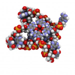
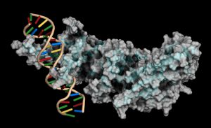
Protein-DNA interactions are fundamental processes in gene regulation in a living cells. These interactions affect a wide variety of cellular processes including DNA replication, repair, and recombination. In vivo methods such as chromatin immunoprecipitation (1) and in vitro electrophoretic mobility shift assays (2) have been used for several years in the characterization of protein-DNA interactions. However, these methods lack the throughput required for answering genome-wide questions and do not measure absolute binding affinities. To address these issues a recent publication (3) presented a high-throughput micro fluidic platform for Quantitative Protein Interaction with DNA (QPID). QPID is an microfluidic-based assay that cam perform up to 4096 parallel measurements on a single device.

The basic elements of each experiment includes oligonucleotides that were synthesized and hybridized to a Cy5-labeled primer and extended using Klenow. All transcription factors that were evaluated contained a 3’HIS and 5’ cMyc tag and were expressed in rabbit reticulocyte coupled transcription and translation reaction (TNT® Coupled Reticulocyte Lysate). Expressed proteins are loaded onto to the QIPD device and immobilized. In the DNA binding assay the fluorescent DNA oligonucleotides are incubated with the immobilized transcription factors and fluorescent images taken. To validate this concept the binding of four different transcription factor complexes to 32 oligonucleotides at 32 different concentrations was characterized in a single experiment. In a second application, the binding of ATF1 and ATF3 to 128 different DNA sequences at different concentrations were analyzed on a single device.

Literature Cited

  1. Ren, B. et al. (2007) Genome-wide mapping of in vivo protein-DNA binding proteins. Science 316, 1497–502.
  2. Garner, M.M. (1981) A gel electrophoresis method for quantifying the binding of proteins to specific DNA regions. Nuc. Acids. Res. 9, 3047-60.
  3. Glick,Y et al. (2016) Integrated microfluidic approach for quantitative high throughput measurements of transcription factor binding affinities. Nuc. Acid Res. 44, e51.

The Cell Line Identity Crisis

29981164-Whistle

If you work with cell lines you may have paid attention to the dramatic headline published last month in the online journal STAT, Thousands of studies used the wrong cells, and journals are doing nothing.” In their column The Watchdogs (“Keeping an eye on misconduct, fraud, and scientific integrity”), Ivan Oransky and Adam Marcus call out the fact that scientists continue to publish research using cell lines that are contaminated or misidentified. Recent estimates have found that the percentage of misidentified cell lines used by scientists is as high as 20 to 36. The blame here is being placed on the peer reviewed journals for not blowing the whistle. The authors call for journals to put some “kind of disclaimer on the thousands of studies affected.”

This is not a new claim. The continuing problem of cell line misidentification, of lack of authentication, has been covered before in various channels. It’s easy to find news publicizing yet another retracted publication. In May 2015 the journal Nature required authors of all submitted manuscripts to confirm the identity of cell lines used in their studies and provide details about the source and testing of their cell lines.

Continue reading “The Cell Line Identity Crisis”

Digging Up More Clues in the History of the Black Death

Bubonic plague victims in a mass grave in 18th century France. By S. Tzortzis [Public domain], via Wikimedia Commons
My last blog post on the Black Death highlighted research that suggested that the reintroduction of Yersinia pestis, the causative agent of the pandemic, originated in Europe during the 14–18th centuries rather than from Asia, the hypothesized origin. In my post, I wrote about my curiosity regarding what an Asian skeleton positive for Y. pestis from that same time period would reveal about the strain or strains that were circulating. Well, a team of researchers has been exploring the issue of strain circulation and an Asian connection, and recently published what they gleaned from additional historic Y. pestis samples in Cell Host & Microbe.

Teeth from 178 individuals in three different locations (two European, one Asian) were screened for Y. pestis infection using the plasminogen activator (pla) gene. Continue reading “Digging Up More Clues in the History of the Black Death”

Magnetic Bacteria Carry Drugs into Tumors

cancer cell

At first glance, the biology of magnetic, underwater-dwelling, oxygen-averse bacteria may seem of little relevance to our most pressing human health problems. But science is full of surprises. A paper published in Nature Nanotechnology presents an inspired use of these bacteria to deliver anti-cancer drugs to tumors, specifically targeting the oxygen-starved regions generated by aggressively proliferating cells.

Continue reading “Magnetic Bacteria Carry Drugs into Tumors”

An Epizootic for the Ages: Revisiting the White-Nose Syndrome Story

Map showing the spread of WNS across North America

In March 2016, two hikers on a trail east of Seattle, WA, found a little brown bat lying on the ground in obviously poor condition. The bat was taken to an animal shelter where it died two days later from White-Nose Syndrome (WNS).

This bat was the first case of WNS found west of the Rocky Mountains. It represented a jump in the spread of WNS, and a troubling one. WNS was first detected in a cave in Albany, New York, and since then it has been moving slowly westward at a rate of about 200 miles per year, according to David Blehert of the United States Geological Survey, the laboratory that confirmed the WNS diagnosis for the Washington bat. Before this year’s discovery outside of Seattle, the westward-most case detected was in eastern Nebraska.

WNS, caused by a cold-loving fungus, Psuedogymnoascus destructans (Pd), can kill 100% of the hibernating bats in a colony, and in the ten years since it has been detected and monitored has killed over 6 million bats in the United States and Canada. As of July 2016, bats infected with the fungus have been found in 29 states and 5 Canadian provinces.

According to Blehert, this is probably the “most significant epizootic of wildlife” ever observed; never before have we seen hibernating mammals specifically affected by a skin fungus. What does that mean? Are we looking at extinction for some bat species? What are the ecological consequences of rapidly losing so many individuals to disease so quickly? And, what, if anything, can be done to combat the disease and help bat populations recover?

Continue reading “An Epizootic for the Ages: Revisiting the White-Nose Syndrome Story”

Easy Automated Genomic DNA Isolation for GMO Testing: From Vision to Reality

29980616-July25-PureFood---Kelly-600x300

The European Union (EU) has a zero tolerance policy for products containing any material from non-authorized genetically modified (GM) crops. Seed entering EU markets may not contain even trace amounts of non-authorized genetically modified material. In 2012, as the global use of GM crops increased, seed testing loads in the EU continued to build. Isolating genomic DNA (gDNA) using traditional manual methods was becoming impractical in the face of increasing amounts of material that required testing. There was a growing need for an automated method to isolate gDNA from seed samples. Working to address this need, a group of scientists from the Bavarian Health and Food Safety Authority collaborated with scientists from Promega Corporation to evaluate the Maxwell® 16 Instrument and the associated chemistry as possible a solution for the testing labs.

Continue reading “Easy Automated Genomic DNA Isolation for GMO Testing: From Vision to Reality”

Shining Light on a Superbug: Clostridium difficile

Antibiotic-resistant bacteria and their potential to cause epidemics with no viable treatment options have been in the news a lot. These “superbugs,” which have acquired genes giving them resistance to common and so-called “last resort” antibiotics, are a huge concern as effective treatment options dwindle. Less attention has been given to an infection that is not just impervious to antibiotics, but is actually enabled by them.

33553646_l

Clostridium difficile Infection (CDI) is one of the most common healthcare-associated infections and a significant global healthcare problem. Clostridium difficile (C. diff), a Gram-positive anaerobic bacterium, is the source of the infection. C. diff spores are very resilient to environmental stressors, such as pH, temperature and even antibiotics, and can be found pretty much everywhere around us, including on most of the food we eat. Ingesting the spores does not usually lead to infection inside the body without also being exposed to antibiotics.

Individuals taking antibiotics are 7-10 times more likely to acquire a CDI. Antibiotics disrupt the normal flora of the intestine, allowing C. diff to compete for resources and flourish. Once exposed to the anaerobic conditions of the human gut, these spores germinate into active cells that embed into the tissue lining the colon. The bacteria are then able to produce the toxins that can cause disease and result in severe damage, or even death.

Continue reading “Shining Light on a Superbug: Clostridium difficile”

microRNA: The Small Molecule with a Big Story

Introduction

miR-34 precursor secondary structure. The colors indicate evolutionary conservation. Ppgardne [GFDL (http://www.gnu.org/copyleft/fdl.html)

RNA molecules have become a hot topic of research. While I was taught about messenger RNA (mRNA), ribosomal RNA (rRNA) and transfer RNA (tRNA), many more varieties have come into the nomenclature after I graduated with my science degrees. Even more interesting, these RNAs do not code for a protein, but instead have a role in regulating gene expression. From long non-coding RNA (lncRNA) to short interfering RNA (siRNA), microRNA (miRNA) and small nucleolar RNA (snoRNA), these classes of RNAs affect protein translation, whether by hindering ribosomal binding, targeting mRNA for degradation or even modifying DNA (e.g., methylation). This post will cover the topic of microRNAs, explaining what they are, how researchers understand their function and role in metabolism, cancer and cardiovascular disease, and some of the challenges in miRNA research.

What are microRNAs? MicroRNAs (miRNAs) are short noncoding RNAs 19–25 nucleotides long that play a role in protein expression by regulating translation initiation and degrading mRNA. miRNAs are coded as genes in DNA and transcribed by RNA polymerase as a primary transcript (pri-miRNA) that is hundreds or thousands of nucleotides long. After processing with a double-stranded RNA-specific nuclease, a 70–100 nucleotide hairpin RNA precursor (pre-miRNA) is generated and transported from the nucleus into the cytoplasm. Once in the cytoplasm, the pre-miRNA is cleaved into an 18- to 24-nucleotide duplex by ribonuclease III (Dicer). This cleaved duplex associates with the RNA-induced silencing complex (RISC), and one strand of the miRNA duplex remains with RISC to become the mature miRNA.

Continue reading “microRNA: The Small Molecule with a Big Story”

How Do Agricultural Landscapes Affect Bee Health?

Honey bee carrying pollen.
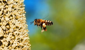
Honey bees are hard-working insects. Their pollination services are in such demand, humans tow hundreds of hives carrying millions of bees around in the back of semitrucks to bring honey bees to various locations such as California almond groves. Humans are also quite partial to the bee colony winter energy storage also known as honey. So while honey bees work hard to collect pollen and nectar from blooming plants and trees and store honey for the winter, humans insist on robbing the colony’s store of delicious sweetener for their own uses. Recent reports of high mortality in honey bee colonies has caused concern in many beekeepers who manage European honey bee apiaries for honey production and pollination services. These severe depletion of honey bee colonies have been attributed to the parasitic mite Varroa destructor in the colony, not only feeding off the larvae and pupae brooding in the colony but also transmitting viruses carried by the mite. Bee nutrition is important for the pollinators especially when overwintering in the hive. Without adequate nutrition, a colony may become weak and succumb to parasite or disease pressure, unable to survive until nectar and pollen are available in the spring. A study was recently published in PLOS ONE that examined how the landscape around Midwestern honeybee hives affected the ability of bees to overwinter and assessed their health by measuring levels of Varroa mites and honey bee viruses. Continue reading “How Do Agricultural Landscapes Affect Bee Health?”

Characterizing Unique Protein: DNA Interactions Using Cell-Free Protein Expression

Molecular model of human telomere DNA

The POT1 protein plays a critical role in telomere protection and telomerase regulation. POT1 binds single-stranded 5′-TTAGGGTTAG-3′ and forms a dimer with the TPP1 protein. Human POT1 contains two Oligonucleotide/Oligosaccharide Binding (OB) fold domains, OB1 and OB2, which make physical contact with the DNA. OB1 recognizes 5′-TTAGGG whereas OB2 binds to the downstream TTAG-3′ (1,2). Several recent studies from other species have shown that some of these proteins are able to recognize a broader variety of DNA ligands than expected (3). A recent reference reexamined the sequence-specificity of the Human POT1 protein (4).
SELEX (Systematic Evolution of Ligands through Exponential Enrichment) was used  to re-examine the DNA-binding specificity of human POT1 (5).

Continue reading “Characterizing Unique Protein: DNA Interactions Using Cell-Free Protein Expression”