Searching for Secrets in Single Cells

There has been a lot of effort recently to perform whole genome sequencing, for humans and other species. The results yield new frontiers of data analysis that offer a lot of promise for groundbreaking scientific discoveries.

One objective of human genome sequencing has been to identify sources of disease and new therapeutic targets. This movement has opened the door to create personalized medicine for cancer, whereby the genetic makeup of an individual’s tumors can be used to determine the most effective drug intervention to administer.

Interest in studying the characteristics unique to individual cells seems obvious when considering the function of healthy cells versus tumor cells, or brain cells compared to heart cells. What has surprised scientists is the realization that two cells in the same tissue can be more different from each other, genetically, than from a cell in another organ.

For example, a small number of brain cells with a specific mutation can lead to some forms of epilepsy while healthy people may also carry cells with these mutations, but too few to cause disease. The lineage of a cell, where it came from and what events shaped its development, ultimately determines what diseases can exist.

Continue reading “Searching for Secrets in Single Cells”

All Aglow in the Ocean Deep

Fascinating bioluminescent organisms floating on dark waters of the ocean. Polychaete tomopteris.

Today’s blog comes to you from the Promega North America Branch Office.

In nature, the ability to “glow” is actually quite common. Bioluminescence, the chemical reaction involving the molecule luciferin, is a useful adaptation for many lifeforms. Fireflies, mushrooms and creatures of the ocean deep use their internal lightshows to cope with a variety of situations. Used for hunting, communicating, ridding cells of oxygen, and simply surviving in the darkness of the ocean depths, bioluminescence is one of nature’s more flashy, and advantageous traits.

In new research published in April in the journal Scientific Reports, MBARI researchers Séverine Martini and Steve Haddock found that three-quarters of all sea animals make their own light.  The study reviewed 17 years of video from Monterey Bay, Calif in oceans that descended to 2.5 miles, to determine the commonality of bioluminescence in the deep waters.

Martini and Haddock’s observations concluded that 76 percent off all observed animals produced some light, including 97 to 99.7 cnidarians (jellyfish), half of fish, and most polychaetes (worms), cephalopods (squid), and crustaceans (shrimp).

Most of us are familiar with the fabled anglerfish, the menacing deep-sea creature known for attracting ignorant prey with a glowing lure attached to their head. As you descend below 200 meters, where light no longer penetrates, you will be surprised at the unexpected color display of the oceans’ sea life. Bioluminescence is not simply an exotic phenomenon, but an important ecological trait that the oceans’ sea creatures have wholeheartedly adopted to cope with complete darkness.

Continue reading “All Aglow in the Ocean Deep”

Revealing Time of Death: The Microbiome Edition

Forensic analysts have long sought precision when determining time of death. While on crime scene investigation television shows, the presence of insects always seems to reveal when a person died, there are many elements to account for, and the probable date may still not be accurate. Insects arrive days after death if at all (e.g., if the body is found indoors or after burial), and the stage of insect activity is influenced by temperature, weather conditions, seasonal variation, geographic location and other factors. All this makes it difficult to estimate the postmortem interval (PMI) of a body discovered an unknown time after death. One way to make estimating PMI less subjective would be to have calibrated molecular markers that are easy to sample and are not altered by environmental variabilities.

Bacterial communities called microbiomes have been frequently in the news. The influence of these microbes encompass living creatures and the environment. Not surprisingly, research has focused on the influence of microbiomes on humans. For example, changes in gut microbiome seem to affect human health. Intriguingly, microbiomes may also be a key to determining time of death. The National Institute of Justice (NIJ) has funded several projects focused on the forensic applications of microbiomes. One focus involves the necrobiome, the community of organisms found on or around decomposing remains. These microbes could be used as an indicator of PMI when investigating human remains. Recent research published in PLOS ONE examined the bacterial communities found in human ears and noses after death and how they changed over time. The researchers were interested in developing an algorithm using the data they collected to estimate of time of death.

Continue reading “Revealing Time of Death: The Microbiome Edition”

In Healthy Eating Less is More: The Science Behind Intermittent Fasting

Mix a love of eating with a desire to live a long, healthy life what do you get? Probably the average 21st-century person looking for a way to continue enjoying food despite insufficient exercise and/or an age-related decline in caloric needs.

Enter intermittent fasting, a topic that has found its way into most news sources, from National Institutes of Health (NIH) and Proceedings of the National Academy of Sciences publications to WebMD and even the popular press. For instance, National Public Radio’s “The Salt” writers have tried and written about their experiences with dietary restriction.

While fasting has enjoyed fad-like popularity over the past several years, it is not new. Fasting, whether purposely not eating or eating a restricted diet, has been practiced for 1,000s of years. What is new is research studies from which we are learning the physiologic effects of fasting and other forms of decreased nutrient intake.

You may have heard the claims that fasting makes people smarter, more focused, and thinner. Researchers today are using cell and animal models, and even human subjects, to measure biochemical responses at the cellular level to restricted nutrient intake and meal timing, in part to prove/disprove such claims (1,2).

Continue reading “In Healthy Eating Less is More: The Science Behind Intermittent Fasting”

Making BRET the Bright Choice for In vivo Imaging: Use of NanoLuc® Luciferase with Fluorescent Protein Acceptors

13305818-cr-da-nanoluc-application_ligund

Live animal in vivo imaging is a common and useful tool for research, but current tools could be better. Two recent papers discuss adaptations of BRET technology combining the brightness of fluorescence with the low background of a bioluminescence reaction to create enhanced in vivo imaging capabilities.

The key is to image photons at wavelengths above 600nm, as lower wavelengths are absorbed by heme-containing proteins (Chu, J., et al., 2016 ). Fluorescent protein use in vivo is limited because the proteins must be excited by an external light source, which generates autofluorescence and has limited penetration due to absorption by tissues. Bioluminescence imaging continues to be a solution, especially firefly luciferase (612nm emission at 37°C), but its use typically requires long image acquisition times. Other luciferases, like NanoLuc, Renilla, and Gaussia, etc. either do not produce enough light or the wavelengths are readily absorbed by tissues, limiting their use to near-surface imaging.

The two papers discussed here illustrate how researchers have combined NanoLuc® luciferase with a fluorescent protein to harness bioluminescent resonance energy transfer (BRET) for brighter in vivo imaging reporters.

Continue reading “Making BRET the Bright Choice for In vivo Imaging: Use of NanoLuc® Luciferase with Fluorescent Protein Acceptors”

Preventing Viral Infection by Blocking Cellular Receptors with a Tethered Antibody

Finding a way to neutralize or block infection by HIV has long been pursued by viral researchers. Various treatments have been developed, driven by the need to find effective drugs to manage HIV in infected individuals. The ultimate goal is to develop a vaccine to prevent HIV from even taking hold in the body. With all of our knowledge about the cellular receptors HIV needs to enter the cell, there has to be a method to prevent a viral particle from binding and being internalized. Many researchers are pursuing neutralizing antibodies to the virus as one method. What about antibodies that target the cellular receptor recognized by the virus? An article published in Proceedings of the National Academy of Sciences, antibodies to cellular receptors for rhinovirus and HIV were tethered to the plasma membrane and tested for the ability to prevent infection.

Continue reading “Preventing Viral Infection by Blocking Cellular Receptors with a Tethered Antibody”

Making a Case for Basic Research Funding

The value of public funding for “basic” versus “applied” research has long been questioned. To address this debate, the authors of a recent report in Science performed a large-scale evaluation of the value of public investment in biomedical research. After analyzing the relationship between the U.S. National Institutes of Health (NIH) grants and private patents, they found that distinguishing research as basic or applied is not useful in determining the productivity of grant funding.

Genetic research at the laboratory

The $30 billion annual budget of the NIH makes it the largest source of life science funding in the world and provides a third of all US biomedical research and development. Although there has long been a strong argument for public investment in scientific research, attacks on the tangible benefits of this research persist. In particular, some opponents argue that “basic” research is too far removed from practical applications to be worthy of investment.

Continue reading “Making a Case for Basic Research Funding”

Making the Switch from FRET to BRET: Applications of NanoLuc® Luciferase with Fluorescent Protein Acceptors for Sensing Cellular Events

A Bioluminescent Alternative

Fluorescence resonance energy transfer (FRET) probes or sensors are commonly used to measure cellular events. The probes typically have a matched pair of fluorescent proteins joined by a ligand-binding or responsive protein domain. Changes in the responsive domain are reflected in conformational changes that either bring the two fluorescent proteins together or drive them apart. The sensors are measured by hitting the most blue-shifted fluorescent protein with its excitation wavelength (donor). The resulting emission is transferred to the most red-shifted fluorescent protein in the pair, and the result is ultimately emission from the red-shifted protein (acceptor).

As pointed out by Aper, S.J.A. et al. below, FRET sensors face challenges of photobleaching, autofluorescence, and, in the case of exciting cyan-excitable donors, phototoxicity. Another challenge to using FRET sensors comes when employing optogenetic regulators to initiate the event you wish to monitor. Optogenetic regulators respond to specific wavelengths and initiate signaling. The challenge comes when the FRET donor excitation overlaps with the optogenetic initiation wavelengths. Researchers have sought to alleviate many of these challenges by exchanging the fluorescent donor for a bioluminescent donor, making bioluminescence resonance energy transfer (BRET) probes. In the three papers described below, the authors chose NanoLuc® Luciferase as the BRET donor due to its extremely bright signal.

Continue reading “Making the Switch from FRET to BRET: Applications of NanoLuc® Luciferase with Fluorescent Protein Acceptors for Sensing Cellular Events”

Probing RGS:Gα Protein Interactions with NanoBiT Assays

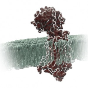
gpcr_in_membrane_on_white2

When I was a post-doc at UT Southwestern, I was fortunate to interact with two Nobel prize winners, Johann Deisenhofer and Fred Gilman.  Johann once helped me move a -80°C freezer into his lab when we lost power in my building. I once replaced my boss at small faculty mixer with a guest speaker and had a drink with Fred Gilman and several other faculty members from around the university. Among the faculty, one professor had a cell phone on his belt, an odd sight in 1995. Fred Gilman asked him what it was and why he had it. It was so his lab could notify him of good results anytime of the day. Fred laughed and told him to get rid of it – if it’s good data, it will survive until morning.

I was reminded of this story when I read a recent paper by Bodle, C.R. et al (1) about the development of a NanoBiT® Complementation Assay (2) to measure interactions of Regulators of G Protein Signaling (RGS) with Gα proteins in cells. (Fred Gilman was the first to isolate a G protein and that led to him being a co-recipient of the Nobel Prize in 1994). The authors created over a dozen NanoBiT Gα:RGS domain pairs and found they could classify different RGS proteins by the speed of the interaction in a cellular context. The interactions were readily reversible with known inhibitors and were suitable for high-throughput screening due to Z’ factors exceeding 0.5. The study paves the way for future work to identify broad spectrum RGS domain:Gα inhibitors and even RGS domain-specific inhibitors. This is the second paper applying NanoBiT Technology to GPCR studies (3).

A Little Background…
A primary function of GPCRs is transmission of extracellular signals across the plasma membrane via coupling with intracellular heterotrimeric G proteins. Upon receptor stimulation, the Gα subunit dissociates from the βγ subunit, initiating the cascade of downstream second messenger pathways that alter transcription (4). The Gα subunits are actually slow GTPases that propagate signals when GTP is bound but shutdown and reassociate with the βγ subunit when GTP is cleaved to GDP. This activation process is known as the GTPase cycle. G proteins are extremely slow GTPases.

Continue reading “Probing RGS:Gα Protein Interactions with NanoBiT Assays”

Writing Scientific Papers: Is There More To This Story?

science & storytelling

The tactic of “telling a good story” is nothing new within the business of selling, marketing and even educating about science. The word itself, “storytelling,” achieved buzzword status a few years ago in the corporate world, so it’s no surprise that it now touches industry scientists.  But the importance of telling a good story within the realm of peer-reviewed scientific papers?  That is something new, and it may impact how scientists write up their results from this point forward.

In a provocative scientific study published in PLOS ONE in December 2016, researchers from the University of Washington showed that “Narrative Style Influences Citation Frequency in Climate Change Science.” Perhaps the results they report are unique to climate change science—an area of science especially susceptible to public perception. But then again, perhaps not. This paper may be worth considering no matter what field of science you call your own.

The authors—Ann Hillier, Ryan Kelly, and Terrie Klinger—used metrics to test their hypothesis that a more narrative style of writing in climate change research papers is more likely to be influential, and they used citation frequency as their measure of influence. A sample of 732 abstracts culled from the climate change literature and published between 2009 and 2010 was analyzed for specific writing parameters. The authors concluded that writing in a more narrative style increases the uptake and influence of articles in this field of science and perhaps in scientific literature across the board.

Continue reading “Writing Scientific Papers: Is There More To This Story?”