Luciferase Immunoprecipitation System Assay (LIPS): Expression of Luciferase Antigen using TNT Transcription/Translation Kit

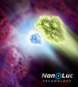
NanoLuc dual reporters
Illustration showing NanoLuc and firefly luciferase reporters.

The luciferase immunoprecipitation system (LIPS) assay is a liquid phase immunoassay allowing high-throughput serological screening of antigen-specific antibodies. The immunoassay involves quantitating serum antibodies by measuring luminescence emitted by the reporter enzyme Renilla luciferase (Rluc) fused to an antigen of interest. The Rluc-antigen fusion protein is recognized by antigen-specific antibodies, and antigen-antibody complexes are captured by protein A/G beads that recognize the Fc region of the IgG antibody (1).

In a recent publication (2), this assay was used to assess the presence of autoantibodies against ATP4A and ATP4B subunits of parietal cells H+, K+-ATPase in patients with atrophic body gastritis and in controls. Continue reading “Luciferase Immunoprecipitation System Assay (LIPS): Expression of Luciferase Antigen using TNT Transcription/Translation Kit”

Ancient Images of Dogs Include Restraints?

This dog is wearing a leash.

You, like me, may occasionally find youself in need of a canine control device. While I’m not a fan of the dog tie out, I do walk dogs on leash—as is required by our county and city government here in Madison, WI.

If you have read Temple Grandin’s books about dogs, you might feel a tug at your heartstrings while enduring a tug on the leash. Aren’t dogs meant to run freely? Don’t we love to watch them run? Are leashes humane?

When walking dogs I feel the need to protect them, but also a desire to let them live like dogs, sniffing, marking and other behaviors that are all limited when the dog is leashed.

When a report in Science last week showed evidence that our ancient ancestors were using leashes 8,000-9,000 years ago I was: 1) surprised; and 2) felt vindicated from self-imposed dog owner guilt.

Continue reading “Ancient Images of Dogs Include Restraints?”

Using CellTiter-Glo® Luminescent Cell Viability Assay to Assess Cell Viability in Cancer Cells Treated with Silver Nanoparticles and DNA-PKcs Inhibitor

Silver nanoparticles (Ag-np) are commonly used in many consumer products, including cosmetics, textiles, electronics and medicine, largely due to their antimicrobial properties. More recently, Ag-np are being used to target and kill cancer cells. It has been known for years that silver nanoparticles (Ag-np) can induce cell death and DNA damage. Studies have also shown that Ag-np inhibit cell proliferation and induce apoptosis in cancer cells. However, cancer cells are able to fight back with DNA repair mechanisms such as non-homologous end joining repair (NHEJ). The NHEJ pathway requires the activation of DNA-dependent protein kinase catalytic subunit (DNA-PKcs), thus DNA-PKcs may protect against the Ag-np-induced DNA damage in cancer cells.

Could inhibition of DNA-PKcs increase the ability of Ag-np to kill cancer cells? In a 2017 study, Lim et al. wanted to test whether inhibition of DNA-PKcs can increase the cytotoxic effect of Ag-np in breast cancer and glioblastoma cell lines. To effectively determine cell viability in these cancer cell lines, the authors used the CellTiter-Glo® Luminescent Cell Viability Assay. The CellTiter-Glo® Assay determines the number of viable cells in culture based on quantitation of ATP, an indicator of metabolically active cells. A major advantage of this assay is its simplicity. This plate-based assay involves adding the single reagent (CellTiter-Glo® Reagent) directly to cells cultured in serum-supplemented medium. This generates a luminescent signal proportional to the amount of ATP present, which is detected using a luminometer. Cell washing, removal of medium and multiple pipetting steps are not required. Another advantage of the CellTiter-Glo® Assay is its high sensitivity. The system detects as few as 15 cells/well in a 384-well format in 10 minutes after adding reagent and mixing, making it ideal for automated high-throughput screening, cell proliferation and cytotoxicity assays.

The authors first confirmed that Ag-np treatment reduced proliferation and induced cell death/DNA damage in two breast cancer cell lines and two glioblastoma cell lines. The cytotoxic effect of Ag-np is specific to cancer cells, as minimal cytotoxicity was observed in non-cancerous human lung fibroblasts used as control. Next, they pre-treated the cancer cells with a DNA-PKcs inhibitor for 1 hour before adding Ag-np. Inhibition of DNA-PKcs increased Ag-np-mediated cell death in all four cancer cell lines. This suggests that DNA-PKcs may be protecting the cells from Ag-np cytotoxicity. The authors further showed that DNA-PKcs may repair Ag-np induced DNA damage by NHEJ and JNK1 pathways. In addition, DNA-PKcs may help recruit DNA repair machinery to damaged telomeres.

This study suggests that a combination of Ag-np treatment and DNA-PKcs inhibition may be a potential strategy to enhance the anticancer effect of Ag-np.

Reference: Hande M.P., et.al. (2017) DNA-dependent protein kinase modulates the anti-cancer properties of silver nanoparticles in human cancer cells. Mutat Res Gen Tox En. 824, 32

Determination of Antibody Mechanism of Action Using IdeS Protease

Monoclonal antibodies (mAbs) have been widely used to eliminate undesired cells via various mechanisms, including antibody-dependent cell-mediated cytotoxicity (ADCC), complement-dependent cytotoxicity (CDC) and programmed cell death (PCD). Unlike the Fc-dependent mechanism of ADCC and CDC, certain antibody–antigen interactions can evoke direct PCD via apoptosis or oncosis. Previously, researchers have reported the specific killing of undifferentiated human embryonic stem cells (hESC) by mAb84 (IgM) via oncosis (1)

In a recent publication (2), a monoclonal antibody (mAb), TAG-A1 (A1), was generated to selectively kill residual undifferentiated human embryonic stem cells (hESC). One of the many experimental tools used to characterize the mechanism of oncosis was the fragmention of the A1 antibody with IdeS and papain.

Learn more about IdeS and IdeZ Protease available from Promega.

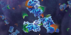
Papain digestion of IgG produces Fab fragments in the presence of reducing agent. F(ab)2 fragments of A1 were produced using IdeS Protease.

The results indicate that both Fab_A1 and F(ab)2_A1 bind to hESC but only F(ab)2_A1 retained hESC killing. Hence bivalency, but not Fc-domain, is essential for A1 killing on hESC.

  1. Choo, A.B. et al. (2008) Selection against undifferentiated human embryonic stem cells by a cytotoxic antibody recognizing podocalyxin-like protein-1. Stem Cells  26, 1454.
  2. Zheng, J.Y. et al. (2017) Excess reactive oxygen species production mediates monoclonal antibody-induced human embryonic stem cell death via oncosis. Cell Death and Differentiation 24, 546–58.

Are you looking for proteases to use in your research?
Explore our portfolio of proteases today.

Hot Wings and Snow Birds: A Study of Genetic Selection in Chickens

African chicken breed Boschvelder. Image copyright ICBH GROUP.

This past summer, I visited the county fair and stopped by the animal barn to look at some of the poultry on display. Specifically, I wanted to see examples of the breeds of chickens available that I may be interested in adding to my flock. Rather than each chicken in their display cage being labeled with a bird’s breed, each cage listed the geographic origin of the chicken within such as Asiatic, Continental or American. This did not benefit my search for potential new members of my flock, but intrigued me enough that I wanted to find out how my flock of 19 hens and pullets would be characterized. Using the classes delineated by the Wisconsin State Fair, my feathered ladies break down to 12 American, 4 English and 3 Continental chickens. There are also classes for Mediterranean and Asiatic (and Other). I live in a part of the United States that gets cold, snowy weather for what seems like six months out of the year, weather that my chickens seem to take in stride. But in other places in the world, heat is the name of the game for the poultry strutting there. In a Genes, Genomics, Genetics publication, Fleming et al. wanted to know if there were genetic differences in Northern European and African chickens that might be caused by their environment.

Continue reading “Hot Wings and Snow Birds: A Study of Genetic Selection in Chickens”

H7N9 Influenza Virus: A Perfect Pathogen?

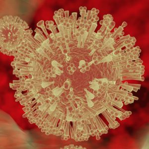
Artist’s rendition of a virus particle.

It’s late October and here in Wisconsin, like many of you, we are experiencing a change of seasons, with the associated drop in temperatures, changes in leaf color and later this week, Halloween.

Another thing that comes with fall is the start of cold and flu season. By “flu”, I mean influenza, caused by avian influenza viruses of the H-N type. Recent research results by teams at UWI-Madison and in Japan, makes the coming influenza season potentially more scary than usual.

In a recent Cell Host & Microbe paper, M. Imai et al. study a seemingly more virulent version of H7N9 avian influenza virus that is startling in its ability to spread from infected to healthy animal models. Based on a current epidemic of H7N9, human-to-human transmission with this strain is increasing.

Continue reading “H7N9 Influenza Virus: A Perfect Pathogen?”

Evaluating DNA Quantity and Quality in FFPE Tumor Samples After Prolonged Storage Using the ProNex® DNA QC Assay

When tumors are surgically removed from cancer patients, the tumor samples are often stored as formalin-fixed and paraffin-embedded (FFPE) tissue blocks. In many cases, tumor samples need to be analyzed several years after diagnosis in order to develop target treatments. But what happens to the DNA after years of storage in FFPE blocks? How well is the DNA preserved?

Scientists in France tried to answer this question in a recent study published in Virchows Arch. The authors extracted DNA from 46 FFPE tumor samples of lung, colon and the urothelial tract, all stored between 4–6 years at room temperature. They then compared the quantity and quality of the DNA to DNA that had been extracted before storage. Using common fluorimetry and qPCR methods, the authors found that the total amount of DNA extracted decreased by half. In addition, the percentage of amplifiable DNA decreased from 56% to only 15% after prolonged storage. Continue reading “Evaluating DNA Quantity and Quality in FFPE Tumor Samples After Prolonged Storage Using the ProNex® DNA QC Assay”

Tick, Tock! The Molecular Basis of Biological Clocks

A long time ago, before the rise of humans, before the first single celled organisms, before the planet even accumulated atmospheric oxygen, Earth was already turning, creating a 24-hour day-night cycle. It’s no surprise, then, that most living things reflect this cycle in their behavior. Certain plants close their leaves at night, others bloom exclusively at certain times of day. Roosters cock-a-doodle-doo every morning, and I’m drowsy by 9:00 pm every night. These behaviors roughly align with the daylight cycles, but internally they are governed by a set of highly conserved molecular circadian rhythms.

Jeffrey Hall, Michael Rosbash and Michael Young were awarded the 2017 Nobel Prize in Physiology/Medicine for their discoveries relating to molecular circadian rhythms. The official statement from the Nobel Committee reads, “…this year’s Nobel laureates isolated a gene that controls the normal daily biological rhythm. They showed that this gene encodes a protein that accumulates in the cell during the night, and is then degraded during the day. [They exposed] the mechanism governing the self-sustaining clockwork inside the cell.” What, then, does this self-sustaining clockwork look like? And how does it affect our daily lives (1)?

Continue reading “Tick, Tock! The Molecular Basis of Biological Clocks”

Analysis of a biosimilar mAb using Mass Spectrometry

Several pharmaceutical companies have biosimilar versions of therapeutic mAbs in development. Biosimilars can promise significant cost savings for patients, but the unavoidable differences
between the original and thencopycat biologic raise questions regarding product interchangeability. Both innovator mAbs and biosimilars are heterogeneous populations of variants characterized by differences in glycosylation,oxidation, deamidation, glycation, and aggregation state. Their heterogeneity could potentially affect target protein binding through the F´ab domain, receptor binding through the Fc domain, and protein aggregation.

As more biosimilar mAbs gain regulatory approval, having clear framework for a rapid characterization of innovator and biosimilar products to identify clinically relevant differences is important. A recent reference (1) applied a comprehensive mass spectrometry (MS)-based strategy using bottom-up, middle-down, and intact strategies. These data were then integrated with ion mobility mass spectrometry (IM-MS) and collision-induced unfolding (CIU) analyses, as well as data from select biophysical techniques and receptor binding assays to comprehensively evaluate biosimilarity between Remicade and Remsima.

The authors observed that the levels of oxidation, deamidation, and mutation of individual amino acids were remarkably similar. they found different levels of C-terminal truncation, soluble protein aggregates, and glycation that all likely have a limited clinical impact.  Importantly, they identified more than 25 glycoforms for each product and observed glycoform population differences.

Overall the use of mass spectrometry-based analysis provides rapid and robust analytical information vital for biosimilar development. They demonstrated the utility of our multiple-attribute monitoring workflow using the model mAbs Remicade and Remsima and have provided a template for analysis of future mAb biosimilars.

1. Pisupati, K. et. al. (2017) A Multidimensional Analytical Comparison of Remicade and the Biosimilar Remsima. Anal. Chem 89, 38–46.

Cytotoxicity Testing of 9,667 Tox21 Compounds using Two Real-Time Assays by Promega

A recent paper in PLOS One demonstrated real-time cytotoxicity profiling of approximately 10,000 chemical compounds in the Tox21 compound library, using two Promega assays, RealTime-Glo™ MT Cell Viability Assay and CellTox™ Green Cytotoxicity Assay. This is exciting to me, a science writer working at Promega; exciting because it’s tricky figuring out how to write about the utility of our products without sounding like an evangelist.

I don’t know about you, but I tend to shut out evangelists and their messages.

Instead of me telling you about real-time viability and cytotoxicity assays from Promega, here is an example of their use in Tox21 chemical compound library research.

What is the Tox21 compound library?
As described in the article by Hsieh, J-H. et al. (2017) in PLOS One:
“The Toxicology in the 21st Century (Tox21) program is a federal collaboration among the National Institutes of Health, including the National Toxicology Program (NTP) at the National Institute of Environmental Health Sciences and the National Center for Advancing Translational Sciences, the Environmental Protection Agency, and the Food and Drug Administration. Tox21 researchers utilize a screening method called high throughput screening (HTS) that uses automated methods to quickly and efficiently test chemicals for activity across a battery of assays that target cellular processes. These assays are useful for rapidly evaluating large numbers of chemicals to provide insight on potential human health effects.”

Continue reading “Cytotoxicity Testing of 9,667 Tox21 Compounds using Two Real-Time Assays by Promega”