Nano, Nano: Tiny Lipid Particles with Big Therapeutic Potential

cell-transfection-viafect-luciferase-assay

Getting DNA or RNA into cells can be a tricky business, and a variety of transfection reagents have been developed over the years to make the process easier. Lipid-based reagents are especially popular because they combine efficient transfection with relatively low toxicity.

When it comes to transfection, it pays to think small. Human cells range in volume from 20–40 µm3 (sperm cells) to as large as 4 million µm3 (mature egg cells, or oocytes). For several decades, transfection reagents have targeted this size range. However, breakthrough research involves leaving the “micro” realm and entering a world that was once the domain only of science fiction: nanotechnology.

Continue reading “Nano, Nano: Tiny Lipid Particles with Big Therapeutic Potential”

A Tale of Two Toxins: the mechanisms of cell death in Clostridium difficile infections

When someone is admitted to a hospital for an illness, the hope is that medical care and treatment will help them them feel better. However, nosocomial infections—infections acquired in a health-care setting—are becoming more prevalent and are associated with an increased mortality rate worldwide. This is largely due to the misuse of antibiotics, allowing some bacteria to become resistant. Furthermore, when an antibiotic wipes out the “good” bacteria that comprise the human microbiome, it leaves a patient vulnerable to opportunistic infections that take advantage of disruptions to the gut microbiota.

One such bacteria, Clostridium difficile, is of growing concern world-wide since it is resistant to many different antibiotics. When a patient is treated with an antibiotic, C. difficile can thrive in the intestinal tract without other bacteria populating the gut. C. difficile infection is the leading cause of antibiotic-associated diarrhea. While symptoms can be mild, aggressive infection can lead to pseudomembranous colitis—a severe inflammation of the colon which can be life-threatening.

C. difficile causes disease by releasing two large toxins, TcdA and TcdB. Understanding the role these toxins play in colonic disease is important for treatment strategies. However, most published research data only report the effects of the toxins independently. A 2016 study demonstrated a method of comparing the toxins side-by-side using the same time points and cell assays to investigate the role each toxin plays in the cell death that leads to disease of the colon. Continue reading “A Tale of Two Toxins: the mechanisms of cell death in Clostridium difficile infections”

Automated Approach for Multiomic Analysis

With the use of a suite of “-omics” technologies you can examine the way in which complex cellular processes work together across all molecular domains (i.e., proteomics, metabolomics, transcriptomics) in a single biological system. Several studies have been published across a wide range of fields illustrating the power of such a unified approach (1,2). Most studies however did not focus on the development of a high-throughput, unified sample preparation approach to complement high-throughput “omic” analytics.

A recent publication by Gutierrez and colleagues presents a simple high-throughput process (SPOT) that has been optimized to provide high-quality specimens for metabolomics, proteomics, and transcriptomics from a common cell culture sample (3). They demonstrate that this approach can process  16−24 samples from a cell pellet to a desalted sample ready for mass spectrometry analysis within 9 hours. They also demonstrated that the combined process did not sacrifice the quality of data when compared to individual sample preparation methods.

Literature Cited

1. Roume, H. (2013) Sequential Isolation of Metabolites, RNA, DNA, and Proteins from the Same Unique Sample. Methods Enzymol. 531, 219−236.
2. Lo, A. W. et al. (2017) ‘Omic’ Approaches to Study Uropathogenic Escherichia Coli Virulence. Trends Microbiol. 25, 729−740.
3. Gutierrez, D. et al. (2018)  An Integrated, High-Throughput Strategy for Multiomic Systems Level Analysis J. Proteome Res.

Are you looking for proteases to use in your research?
Explore our portfolio of proteases today.

From Drug Screening to Agriculture to Cardiac Development, A Dual-Luciferase Reporter Brings You the Story

Today’s blog was written by guest blogger Katarzyna Dubiel, marketing intern in Cellular Analysis and Proteomics. Last updated 02/12/2021

Reporter gene assays have been critical for the study of a wide-range of biological questions, from regulation of gene expression to cellular signaling. While reporter gene assays constitute a large group of technologies, here we highlight the diversity of new discoveries enabled by highly quantitative and easily measured bioluminescent luciferase-based reporter assays. Below are our top picks of exciting research discoveries involving the Dual-Luciferase Reporter Assay format using firefly and Renilla luciferases.

Continue reading “From Drug Screening to Agriculture to Cardiac Development, A Dual-Luciferase Reporter Brings You the Story”

How Autophagy Feeds Cancer’s Need for Metabolites

Illustration of energy metablism in cell.

Metabolism underpins numerous cellular processes. Without it, cells would not grow, divide, synthesize or secrete. Another pathway, autophagy, degrades unwanted cellular materials, helping to maintain cell health. With these opposing roles, is there a connection between autophagy and metabolism? As it turns out, the answer is yes. Because molecules degraded by autophagy are recycled and fed into metabolism pathways as precursor compounds. There are interesting implications as a result of this connection, ones that affect cancer cells as described in a recent Cell Metabolism review article.

Autophagic flux, the process by which molecules and organelles are directed to the autophagosome, fuse with the lysosome and are degraded, involves a selective process that determines the cargo carried within the autophagosome. Autophagy-related genes (ATGs) direct the process and particular receptor proteins bind the cargo. What is interesting about the connection among cancer, autophagy and metabolism is the complexity of the role that autophagy plays in cancer. While autophagy was thought to act in a more tumor suppressive manner as shown when one copy of an ATG6 analogous gene in mice was deleted and the other left unaltered, and malignant tumors developed, but in mice mosaic for ATG5 deletions, the inhibition of autophagy resulted in benign tumors in the liver. This latter experiment suggested autophagy was needed for cancer progression, a hypothesis reinforced by the lack of ATG mutations in human cancers.

Continue reading “How Autophagy Feeds Cancer’s Need for Metabolites”

Autophagy: The Poem

Roberta A. Gottlieb, MD, is the Director of Molecular Cardiobiology at Cedar-Sinai, a nonprofit academic healthcare organization. She is interested in the role of autophagy in myocardial ischemia, a kind of heart disease in which blood flow to the heart is blocked. (Studies have shown that autophagy is upregulated during myocardial ischemia, but why this happens is not entirely clear.) Her ultimate goal is to understand and mitigate ischemic injury, with the hope of developing therapeutics for humans.

And—she’s a poet.

Continue reading “Autophagy: The Poem”

Using Environmental DNA to Find Sharks

Sharks are often known as one of the fiercest predators in the ocean. Yet they are also one of the most threatened marine species—largely because they are hunted by humans for their highly valuable fins. How do we know they are being threatened?

Traditionally, researchers have estimated the population and diversity of sharks using two methods. One is by sending divers into the water to count the number of sharks and record which species they see. Another is to set up baited cameras in different locations to record the sharks that come near. When fewer species of sharks are examined using these methods, we conclude that there is a decrease in diversity and an increase in what’s called “dark diversity”.

Continue reading “Using Environmental DNA to Find Sharks”

High-Throughput Drug Screening Using 3D Cell Cultures

For a long time, the drug industry has relied on flat 2D cell cultures grown on a plate to screen for potential drugs. However, 2D models do not accurately reflect the native environment of cells in vivo. 3D cell cultures, on the other hand, better represent the numerous cell-cell and cell-matrix interactions and hypoxic conditions that have a profound effect on the behavior of cells. In a 2018 study published in Oncogene, Kota et al. developed a high-throughput 3D spheroid-based screening assay to identify drug candidates that target RAS proteins.

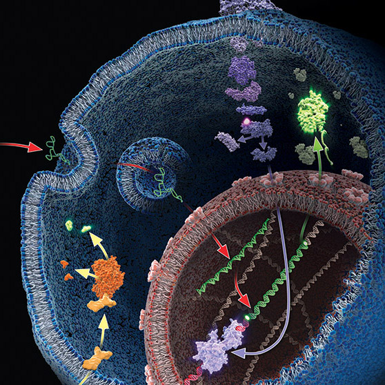
RAS proteins are GTPases that transmit extracellular signals into cellular signaling pathways, which could activate cell proliferation, differentiation and survival mechanisms. Oncogene mutation in the three human RAS genes (HRAS, NRAS and KRAS) are found in 30% of all cancers, making RAS proteins the most common oncogene. In fact, mutations in KRAS are found in >90% of pancreatic cancers. Despite the prevalence of RAS mutations, targeting RAS proteins with drugs is extremely challenging due to the complex nature of the protein.

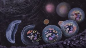
The authors in this study wanted to test a new approach using a 3D spheroid-based screening assay to find drugs that target RAS proteins. They first harvested 2D monolayer cultures of pancreatic epithelial tumor cells that express either wild-type KRAS or mutant oncogenic KRAS, and tested their ability to form 3D spheroids. They confirmed spheroid growth using the CellTiter-Glo® 3D Cell Viability Assay with linearity of detection in the range of 1,000–10,000 cells seeded.

The 3D spheroids were then treated with a library of 1,280 known drugs. From the high-throughput screen, they identified one compound with the greatest selective inhibition against oncogenic KRAS. The compound is called Proscillaridin A, a cardiac glycoside that is known for treating congestive heart failure and cardiac arrhythmia. In 3D spheroids, Proscillardin A inhibited oncogenic KRAS at a >90% inhibition rate, with <10% inhibition of wild-type KRAS. In 2D cultures, however, there was no selective inhibition of oncogenic KRAS (inhibition rates for both oncogenic and wild-type KRAS were about 50%). This means that Proscillaridin A would not have been identified as a candidate if the screen was done using only 2D cultures.

Next, the authors wanted to determine how Proscillaridin A impacts tumor cell viability. Could it induce apoptosis in tumor cells? To test this, they used the RealTime-Glo™ Annexin V Apoptosis Assay. This bioluminescent assay is able to detect apoptosis in real time, based on the exposure of phosphatidylserine on the outer leaflet of the cell membrane when apoptosis occurs. Using this assay, they found that Proscillaridin A induced apoptosis at earlier time points and higher rates in 3D spheroids expressing oncogenic KRAS compared with wild-type KRAS. In 2D cultures, there was no difference in the rate of apoptosis.

This study shows that high-throughput screening in 3D spheroids can identify potential drugs that would not have been discovered in a 2D format. This provides hope for finding drugs against difficult target proteins such as RAS.

Reference: Kota S., et al. (2018) A novel three-dimensional high-throughput screening approach identifies inducers of a mutant KRAS selective lethal phenotype. Oncogene. Epub ahead of print.

Bacteria and Viruses as Cancer Treatments

Over a hundred years ago William B Coley, the “Father of Immunotherapy”, discovered that injection of bacteria or bacterial toxins into tumors could cause those tumors to shrink. The introduction of bacteria had the side-effect of stimulating the immune system to attack the tumor. The field of cancer immunotherapy research—which today includes many different approaches for generating anti-tumor immune responses—originated with these early experiments.

Use of bacteria is one way to stimulate the immune system to attack cancer cells, others include use of cytokines, immune checkpoint blockades and vaccines. This Nature animation provides a simple overview of these methods.

Continue reading “Bacteria and Viruses as Cancer Treatments”

Finding Chinks in the Armor: Cancer’s Need for Metabolites

Illustration of energy metablism in cell.

Cancer has been studied for decades by scientists trying to find a vulnerability to exploit and testing compounds to develop as potential drugs. As the “Emperor of All Maladies”, cancer has proven itself to be a wily beast with many varieties of genetic mutations for eluding cellular control, tireless in its ability to divide and spread. In the end, a cancer cell is still a cell and subject to its environment even though cancer does not play by the same rules as the normal cells that exist around it. To be able to grow, a cell needs access to metabolites, molecules needed for building the materials and machinery needed by the cell to function and divide. These requirements also offer potential pathways to target for halting cancer growth and spread.

All cells use glucose to generate ATP, but normal and cancer cells differ in how glucose is converted to ATP. Most cells use glucose in oxidative phosphorylation, but cancer cells use aerobic glycolysis, converting glucose to lactate without oxygen. This Warburg effect (glucose converted to lactate) is a hallmark of cancer cells as they take up glucose at a much higher rate than normal cells. Blocking glucose uptake is one way to target cancer cells. While 2-deoxyglucose (2DG) has been shown to slow glucose uptake in vitro, the compound proved toxic in clinical trials and lower dosages do not seem to be an effective treatment against cancer. While not an ideal drug target, glucose uptake has been helpful in monitoring cancer response to therapies via fluorodeoxyglucose positron emission tomography (FDG-PET).

Continue reading “Finding Chinks in the Armor: Cancer’s Need for Metabolites”