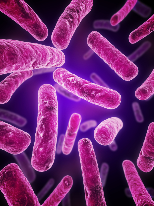
Think about the last time you gave a presentation. The feeling of having “butterflies in your stomach”. Or when you meet someone for the first time, that “gut feeling” of whether you two will get along. In our day-to-day lives, we often associate what happens in the gut with what goes on in our brain. In fact, scientific evidence suggests that our gut and our brain frequently communicate—through gut microbes. Apparently, the existence of trillions of bacteria and eukaryotes in our gut is not only crucial for our physical health, they may also be important for our mental health.
Yersinia pestis. See page for author [Public domain], via Wikimedia Commons
In recent years, scientists have been able to refine their molecular tools to resurrect ancient DNA from human graves and determine that yes, Yersinia pestis was the causative agent for the Black Death in the 14th century and the Plague of Justinian in the 6th century. As more and more human graves have been uncovered, their DNA has revealed many secrets that scientists even ten years ago were unable to discover. With the ability to sequence entire genomes of bacteria that died with their hosts hundreds and even thousands of years ago, researchers are exploring the rise and possible spread of Y. pestis. Each new member sequence adds to the Y. pestis family tree, pinpointing the origin of this bacteria as it diverged from its ancestor Y. pseudotuberculosis. Peering into the past, scientists have been able to track down a strain of Y. pestis from individuals in a Swedish passage grave that is basal to known strains and that the authors of a Cell article suggest has interesting implications.
This pathogenic journey into history started by analyzing ancient DNA data sets from the teeth of individuals present in a communal passage grave in Gökhem parish, located in western Sweden, for any disease-causing microbial sequences that might be present. Y. pestis was flagged in one 20-year-old female dated 4,867–5,040 years ago. The bacterial sequences from this individual, named Gok2, were more closely aligned with Y. pestis than the Y. pseudotuberculosis reference genome.
Ebola virus (EBOV) and Marburg virus (MARV) are two closely-related viruses in the family Filoviridae. Filoviruses are often pathogenic, causing hemorrhagic fever disease in human hosts. The Ebola outbreak of 2014 caught the world by surprise by spreading so quickly and severely that public health organizations were unprepared. The devastating outcome was a total of over 11,000 deaths by the time the outbreak ended in 2016. Research that provides further understanding of filoviruses and their potential for transmission is important in preventing future outbreaks from occurring. But what if the outbreak comes from a virus we’ve never seen before?
Měnglà virus was discovered among filoviruses isolated from Old World fruit bats (Rousettus)
All in the viral family
A recent study published in the journal Nature Microbiology provides evidence of a newly identified filovirus species. Using serum samples taken from bats, a well-known host for filoviruses, Yang et al. isolated and identified viral RNA for an unclassified viral genome sequence using next generation sequencing analysis. This new virus genome sequence was organized with the same open reading frames as other filoviruses, encoding for nucleoprotein (NP), viral protein 35 (VP35), VP40, glycoprotein (GP), VP30, VP24, and RNA-dependent RNA polymerase (L). This new genome sequence shared up to 54% of the nucleotide sequences for the filovirus species Lloviu virus (LLOV), EBOV and MARV, with MARV being the most similar. Their analysis suggested that this novel virus should be classified within the Filoviridae family tree as a separate genus, Dianlovirus, and was named Měnglà virus (MLAV).
Redundancy equips us to survive. We have more than one lung or one kidney for a reason—if one organ in a pair gets damaged, we can still manage if the other is functional. At the cellular level, we have two copies of each chromosome in every non-germline cell. Each copy was inherited originally from a single sperm and ovum, which are “haploid” cells. Consequently, there are two copies of any given gene in non-germline “diploid” cells. In many cases, should one copy of a gene be damaged, the cell can still survive with the other, functional copy of a gene. In plants, this redundancy is common, and many plants exhibit polyploidy. In an extreme example of polyploidy, the large (by bacterial standards) but otherwise unassuming species Epulopiscium contains tens of thousands of copies of its genome.
In almost every environment on earth, such as soil, human
skin and gut, there lives a whole community of microbes—sometimes up to
hundreds of species. It may seem like they all flourish in peace. But just like
you may have friendly or hostile interactions with your neighbors, the
different bacterial species interact in various ways. They may cooperate,
compete or, sometimes, even kill each other. The interaction is complicated,
and scientists have struggled to understand the nature of these microbiome
interactions. How do microbiomes assemble and maintain stability? How do the
interactions among different species affect gene expression?
A. gossypii on cotton leaf. Image credit: Clemson University – USDA Cooperative Extension Slide Series, , United States [CC BY 3.0
The extensive and repetitive use of neonicotinoids has led to the development of resistance in several insect species including, the cotton aphid, A. gossypii. A. gossypii is a widely distributed pest that affects watermelons, cucumbers, pumpkin, cotton, and citrus crops, among others, making it one of the most economically important agricultural pests known. Thiamethoxam is a neonicotinoid insecticide that irreversibly binds to the nicotinic acetylcholine receptors (nAChRs) of cells in the nervous system and interferes with the transmission of nerve impulses in insects (1).
To further understand the mechanisms of resistance to thiamethoxam and other neonicotinoids, Wu et al. recently investigated (2) expression changes in the transcripts of P450 in thiamethoxam-susceptible and thiamethoxam-resistant cotton aphid strains. Nine P450 genes were significantly overexpressed in the resistant strain (especially CYP6CY14). The involvement of overexpressed P450s was examined through RNA interference (RNAi) introduced via artificial diet and dsRNA feeding.
Have you ever had a day where you feel exceptionally good? As intake on the world kind of good? You feel so much better than the previous couple of days that you stop to wonder why.
Then it dawns on you.
The sun is out. It’s been cloudy for the past week but now—SUNSHINE.
You go out to lunch or for a walk just to take in those rays. Sure, it feels warmer than your darkened office space, but it’s the light rather than warmth that’s making a difference.
You purposely don’t wear sunglasses and it feels like the light is coming in through your eyes and massaging that part of your brain that is your happy zone. Are you imagining it or is the sun really affecting how you feel?
In a study reported in the September 2018 issue of Cell we learn that this is not a figment of your or my imagination (1). There is, in fact, a type of retinal cell that transports sunlight directly to the part of our brains that affects mood.
Eyes and the Body’s Master Clock
Circadian rhythms are innate time-keeping functions found in all multicellular organisms. This subject of the 2017 Nobel prize in Physiology or Medicine, circadian rhythms are fueled by daily light-dark cycles and are critical to the function of neurologic, immune, musculoskeletal and cardiac tissues (2). Nearly every mammalian cell is affected by circadian rhythms.
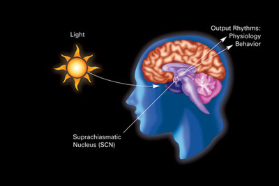
The human body has a circadian master clock, the suprachiasmatic nucleus or SCN. The SCN is a highly innervated tissue located in the hypothalamus (see image). It is connected directly to the retina by the optic nerve, and thus is influenced by external light and dark.
Light enters the eyes and affects the SCN (physiologic effects), and as discussed in recent research, Fernandez et al. here, the perihabenular nucleus (behavioral effects). (Image in public domain.)
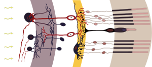
The retina of the eye is the light-gathering instrument for this organ. Historically, it’s been understood that the retina is composed of two cell types, rods and cones, that function in transmitting light and images to the optic nerve, which sends those signals to the brain.
Some parts of the retina. Light enters the eye (from left) and passes through to the rods and cones. Here a chemical change converts the light to nerve signals. Image-based on drawing by Ramón y Cajal, 1911 and licensed under Wikimedia commons.
Studies by Hattar et al. in the early 2000s identified another cell found in the retina, the melanopsin-containing intrinsically photoactive retinal ganglion cells (ipRGCs) as the transmitter of circadian light signals (3). Through this direct connection to the SCN, the circadian master clock, the ipRGCs can influence a wide range of light-dependent functions independent of image processing (4).
Now Fernandez et al. have identified multiple types of ipRGCs. They showed that ipRGCs that mediate the effects of light on learning work via the SCN, while the pathway for light influencing emotions is different.
They discovered a new target of ipRGC cells, the perihabenular nucleus (PHb). The PHb is a newly recognized thalamic region of the brain. The authors showed that the connection between light and mood is regulated by ipRGCs through the PHb versus the SCN. They show that the PHb is integrated into other mood-regulating centers of the thalamic region.
In Conclusion
Daylight, and lack thereof, does affect both our mood and our ability to learn. In this 2018 report, we have learned that the pathways for these effects are distinct, and gain an understanding of a new thalamic region by which the light and mood actions occur. This information could influence the development of better drugs and/or therapies for major depressive disorders.
For those of us with seasonal affective disorder, the evidence is undeniable—lack of light can cause issues, from sleep-wake problems, to mood and learning issues.
And while we can’t create sunshine, a special lamp or lightbox may help to gain some full-spectrum light. To learn more about how to choose such a lamp and when to use it, see this Mayo clinic article for details.
We have published 130 blogs here at Promega this year (not including this one). I diligently reviewed every single one and compiled a list of the best 8.5%, then asked my coworkers to vote on the top 5 out of that subset. Here are their picks:
Glucose is an energy metabolite necessary for cellular survival and growth whether or not the cell is part of a tumor. Not only do cancer cells switch from oxidative phosphorylation to aerobic glycolysis (the Warburg effect) to gain more glucose, a hallmark of cancer, but they also increase the amount of glucose taken up from the surrounding extracellular space. However, the lack of glucose can have a negative effect on cells, causing them to become apoptotic in the absence of this metabolite. Cancer cells have methods to get around the requirement for glucose, including upregulating glucose transporters to improve access to the energy metabolite. In this Redox Biology article, researchers describe how activating androgen receptor in response to a lack of glucose affects the amount of GLUT1 expressed on prostate cancer cells, making the cells resistant to glucose deprivation.
To set the stage, two prostate cancer cell lines, LNCaP, an androgen-sensitive cell line, and LNCaP-R, an androgen-insensitive cell line, were deprived of glucose. Both cell lines showed signs of cell death, but LNCaP-R cells died in greater numbers. To probe how LNCaP cells died, several inhibitors (a pan-caspase inhibitor, two necroptosis inhibitors and a ferroptosis inhibitor) were added but did not change the way the cells died. However, an autophagy inhibitor enhanced cell death, suggesting the cells were necrotic not apoptotic. Teasing apart if the necrosis of LNCaP cells was due to glucose availability or merely disrupted glycolysis, the glucose analog 2DG was added to the medium with glucose. The cells survived when treated with 2DG, suggesting it was the absence of glucose that induced necrosis. When LNCaP cells were cultivated in medium that replaced glucose with mannose or fructose, the cells survived, another point in favor of sugar depletion causing cell death.
The vagal nerve could serve as conduit for transit of alpha-synuclein from appendix to brain.
Since about 2000 we’ve learned a lot about the bacteria in our guts. We’ve learned that the right bacterial communities in our gastrointestinal system can make us feel better, think better and even help avoid obesity (1). My colleague Isobel has previously blogged about how certain gut bacteria can improve immunotherapy outcomes.
Conversely, the wrong bacteria in our guts can have negative consequences on health and cognition.
Along the way we’ve learned that gut bacterial flora can be influenced by what we eat, certain medications like antibiotics, and even stressful events. We now know that fermented foods like yogurt, sauerkraut, kombucha and that horrible-smelling stuff (kimchi) that another colleague eats are happy food for the good gut bacteria.
XWe use cookies and similar technologies to make our website work, run analytics, improve our website, and show you personalized content and advertising. Some of these cookies are essential for our website to work. For others, we won’t set them unless you accept them. To learn more about our approach to Privacy we invite you to Read More
By clicking “Accept All”, you consent to the use of ALL the cookies. However you may visit Cookie Settings to provide a controlled consent.
We use cookies and similar technologies to make our website work, run analytics, improve our website, and show you personalized content and advertising. Some of these cookies are essential for our website to work. For others, we won’t set them unless you accept them. To find out more about cookies and how to manage cookies, read our Cookie Policy.
If you are located in the EEA, the United Kingdom, or Switzerland, you can change your settings at any time by clicking Manage Cookie Consent in the footer of our website.
Necessary cookies are absolutely essential for the website to function properly. These cookies ensure basic functionalities and security features of the website, anonymously.
Cookie
Duration
Description
cookielawinfo-checbox-analytics
11 months
This cookie is set by GDPR Cookie Consent plugin. The cookie is used to store the user consent for the cookies in the category "Analytics".
cookielawinfo-checbox-functional
11 months
The cookie is set by GDPR cookie consent to record the user consent for the cookies in the category "Functional".
cookielawinfo-checbox-others
11 months
This cookie is set by GDPR Cookie Consent plugin. The cookie is used to store the user consent for the cookies in the category "Other.
cookielawinfo-checkbox-advertisement
1 year
The cookie is set by GDPR cookie consent to record the user consent for the cookies in the category "Advertisement".
cookielawinfo-checkbox-necessary
11 months
This cookie is set by GDPR Cookie Consent plugin. The cookies is used to store the user consent for the cookies in the category "Necessary".
cookielawinfo-checkbox-performance
11 months
This cookie is set by GDPR Cookie Consent plugin. The cookie is used to store the user consent for the cookies in the category "Performance".
gdpr_status
6 months 2 days
This cookie is set by the provider Media.net. This cookie is used to check the status whether the user has accepted the cookie consent box. It also helps in not showing the cookie consent box upon re-entry to the website.
lang
This cookie is used to store the language preferences of a user to serve up content in that stored language the next time user visit the website.
viewed_cookie_policy
11 months
The cookie is set by the GDPR Cookie Consent plugin and is used to store whether or not user has consented to the use of cookies. It does not store any personal data.
Analytical cookies are used to understand how visitors interact with the website. These cookies help provide information on metrics the number of visitors, bounce rate, traffic source, etc.
Cookie
Duration
Description
SC_ANALYTICS_GLOBAL_COOKIE
10 years
This cookie is associated with Sitecore content and personalization. This cookie is used to identify the repeat visit from a single user. Sitecore will send a persistent session cookie to the web client.
vuid
2 years
This domain of this cookie is owned by Vimeo. This cookie is used by vimeo to collect tracking information. It sets a unique ID to embed videos to the website.
WMF-Last-Access
1 month 18 hours 24 minutes
This cookie is used to calculate unique devices accessing the website.
_ga
2 years
This cookie is installed by Google Analytics. The cookie is used to calculate visitor, session, campaign data and keep track of site usage for the site's analytics report. The cookies store information anonymously and assign a randomly generated number to identify unique visitors.
_gid
1 day
This cookie is installed by Google Analytics. The cookie is used to store information of how visitors use a website and helps in creating an analytics report of how the website is doing. The data collected including the number visitors, the source where they have come from, and the pages visted in an anonymous form.
Advertisement cookies are used to provide visitors with relevant ads and marketing campaigns. These cookies track visitors across websites and collect information to provide customized ads.
Cookie
Duration
Description
IDE
1 year 24 days
Used by Google DoubleClick and stores information about how the user uses the website and any other advertisement before visiting the website. This is used to present users with ads that are relevant to them according to the user profile.
test_cookie
15 minutes
This cookie is set by doubleclick.net. The purpose of the cookie is to determine if the user's browser supports cookies.
VISITOR_INFO1_LIVE
5 months 27 days
This cookie is set by Youtube. Used to track the information of the embedded YouTube videos on a website.
Performance cookies are used to understand and analyze the key performance indexes of the website which helps in delivering a better user experience for the visitors.
Cookie
Duration
Description
YSC
session
This cookies is set by Youtube and is used to track the views of embedded videos.
_gat_UA-62336821-1
1 minute
This is a pattern type cookie set by Google Analytics, where the pattern element on the name contains the unique identity number of the account or website it relates to. It appears to be a variation of the _gat cookie which is used to limit the amount of data recorded by Google on high traffic volume websites.


![A. gossypii on cotton leaf. Image credit: Clemson University - USDA Cooperative Extension Slide Series, , United States [CC BY 3.0 (https://creativecommons.org/licenses/by/3.0)], via Wikimedia Commons](https://www.promegaconnections.com/wp-content/uploads/2019/01/Aphis_gossypii04-300x290.jpg)