Nucleic Acid Quantitation by UV Absorbance: Not for NGS

schematic diagram of UV-Vis Absorbance Method
For UV-Vis Spectrophotometry, light is split into its component wavelengths and directed through a solution. Molecules in the solution absorb specific wavelengths of light.

This is the second in a series of four blogs about Quantitation for NGS is written by guest blogger Adam Blatter, Product Specialist in Integrated Solutions at Promega.

Perhaps the most ubiquitous quantitation method is UV-spectrophotometry (also called absorbance spectroscopy). This technique takes advantage of the Beer-Lambert Law: an observation that many compounds absorb UV-Visible light at unique wavelengths, and that for a fixed path length the absorbance of a solution is directly proportional to the concentration of the absorbing species. DNA, for example has a peak absorbance at 260nm (A260nm).

This method is user friendly, quick and easy. But, it has significant limitations, especially when quantitating samples for NGS applications.

Continue reading “Nucleic Acid Quantitation by UV Absorbance: Not for NGS”

When Every Step Counts: Quantitation for NGS

13170MA-800x277This series of blogs about Quantitation for NGS is written by guest blogger Adam Blatter, Product Specialist in Integrated Solutions at Promega.

As sequencing technology races toward ever cheaper, faster and more accurate ways to read entire genomes, we find ourselves able to study biological systems at a level never before possible. From basic science to translational research, massively parallel sequencing (also known as next-generation sequencing or NGS) has opened up new avenues of inquiry in genomics, oncology and ecology.

Many commercial sequencing platforms have been established (e.g., Illumina, IonTorrent, 454, PacBio), and new technologies are developed every day to enable new and unique applications. However, all of these platforms and technologies work off the same general principle: nucleic acid must be extracted from a sample, arranged into platform-specific library constructs, and loaded into the sequencer. Regardless of the sample type or the platform used, every step throughout this workflow is critical for successful results. An often overlooked part of the NGS workflow is sample quantitation. Here we are presenting the first in a series of four short blogs about the critical step of quantitation in NGS workflows.

Sample input is critical to NGS in terms of both quality and quantity. Knowing how much DNA you have, often in nanogram quantities, can make the difference between success and failure. There are several key points in the NGS workflow where sample quantitation is important before you can proceed:

  • After initial nucleic acid extraction from the sample matrix and before proceeding with library preparation
  • Post-library preparation when pooling barcoded libraries for multiplexing
  • Final pooled library quantitation immediately before loading for sequencing

There are several common methods for quantitating nucleic acids: UV-spectroscopy, Fluorescence spectoscopy, real-time quantitative PCR (qPCR). Because of inherent differences in sensitivity, specificity, time and cost, each of these techniques pose certain advantages and disadvantages with respect to the specific sample you are quantitating. Our next three blogs will discuss each of these methods against the backdrop of quantitating samples for NGS applications.


Read Part 2: Nucleic Acid Quantitation by UV Absorbance: Not for NGS

Read Part 3: Fluorescence Dye-Based Quantitation: Sensitive and Specific for NGS Applications

Read Part 4: Real-Time (Quantitative) qPCR for Quantitating Library Prep before NGS

DNA Purification from Plants: Not All Methods are Equal

pestle and mortar with leavesIsolating DNA from plant tissues is difficult for many reasons.  Unlike animal cells, plant cells have rigid cell walls, often made of tough fibrous material, and contain proteins and enzymes and other compounds such as polysaccharides and polyphenols that play a role in different cellular processes. These compounds can interfere with DNA isolation as well as downstream applications such as PCR.  For these reasons, DNA isolation methods that are used successfully for other sample types may not work well to isolate DNA from plant material. Continue reading “DNA Purification from Plants: Not All Methods are Equal”

Freedom to Focus: Maxwell® Rapid Sample Concentrator

Wish I had one of these when I was at the lab bench...
Wish I had one of these when I was at the lab bench…

Back in the dark ages, when I was a graduate student, my idea of “automated” plasmid DNA extraction involved performing home-brew, “toothpick preps” in “strip tubes” or , if I was really feeling ambitious, a 96-well plate.

I would get just enough DNA to check for the presence of an insert, but the quality of the DNA was too low and the quantity too small to even consider using it for any other downstream experiments like amplification.

And increased throughput for other nucleic acid extraction needs? Nope. If I wanted genomic DNA, RNA or high-quality plasmid DNA, I spent time with columns and tubes, giving each sample my undivided individual attention.

Remember cesium chloride preps for RNA isolation? Even with the advent of column purification, which greatly simplified and standardized my protocols, nucleic acid purification was still a manual task that required a lot of time and effort to get the high-quality product I needed.

Doing the experiments that would answer the questions that I really wanted to ask (those “downstream experiments”), meant spending time at the bench performing careful (if tedious) work to isolate and clean up the highest quality nucleic acid possible. Even then inconsistency in sample prep could wreak havoc on downstream work.

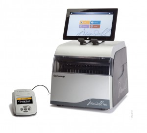
Fortunately, for the modern scientist, personal, bench top automation, has progressed far beyond the toothpick and the strip tube to quality-tested, reliable nucleic acid extraction platforms like the Maxwell® Rapid Sample Concentrator (RSC).

The Maxwell® RSC improves sample preparation consistency, eliminating variability associated with manual handling, and your downstream results will reflect this consistency.  With the RSC you can extract DNA or RNA from up to 16 samples in approximately 1 hour and viral total nucleic acids in less than an hour.

The instrument is easy to use: simply load the sample, push a button and walk away. Cross contamination is minimized and the instrument is supported by the Promega technical support and service you have come to trust over the past 35 years. 


Want to know more about how the Maxwell® RSC can give you the freedom to focus on the work that interests you the most? To learn more, click here.


Dealing with PCR Inhibitors

Inhibition

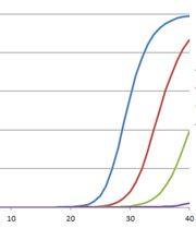
The polymerase chain reaction (PCR) has revolutionized modern biology as a quick and easy way to generate amazing amounts of genomic data. However, when PCR doesn’t work, it can be frustrating. At these times, PCR and reverse transcription PCR (RT-PCR) inhibitors seem to be everywhere: They lie dormant in your starting material and can co-purify with the template of interest, and they can be introduced during sample handling or reaction setup. The effects of these inhibitors can range from partial inhibition and underestimation of the target nucleic acid amount to complete amplification failure. What is a scientist to do?

Continue reading “Dealing with PCR Inhibitors”

Methods for Quantitating Your Nucleic Acid Sample

Nucleic acid quanitation webinar

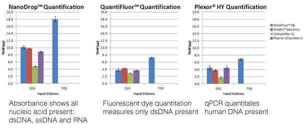
For most molecular biology applications, knowing the amount of nucleic acid present in your purified sample is important. However, one quantitation method might serve better than another, depending on your situation, or you may need to weigh the benefits of a second method to assess the information from the first. Our webinar “To NanoDrop® or Not to NanoDrop®: Choosing the Most Appropriate Method for Nucleic Acid Quantitation” given by Doug Wieczorek, one of our Applications Scientists, discussed three methods for quantitating nucleic acid and outlined their strengths and weaknesses.

Continue reading “Methods for Quantitating Your Nucleic Acid Sample”

Don’t Let Ribonucleases Ruin Your Week(end): Establish a Ribonuclease-free Environment

buffers_image1_274x218

My very first job in science was in a lab that worked exclusively with RNA, and it was only after I moved on to a different job that I learned just how much different the world of DNA research is from that of RNA. When working with DNA, for example, you rarely if ever have the sample you have labored over reduced to a fuzzy blur at the bottom of a gel because it has been degraded beyond rescue. With RNA, unfortunately, this happens all too frequently. In fact, a labmate of mine once put up a poll on the door to our lab asking if it was better to discover that your RNA sample was degraded on a Monday or a Friday.

The culprits in this scenario are Ribonucleases (RNases). They are everywhere. They are incredibly stable and difficult to inactivate. And, if you work with RNA, they are your enemy. Take heart though, they can be defeated if you follow some pretty simple steps.

Continue reading “Don’t Let Ribonucleases Ruin Your Week(end): Establish a Ribonuclease-free Environment”

Safety First (and Sensitivity too!): Diamond™ Nucleic Acid Dye

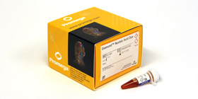
product photo for diamond™ nucleic acid dye

Gel electrophoresis and gel staining are common lab tasks that you may not think too much about.  It’s a fairly routine part of your day…purify DNA or RNA, check it on a gel.  As you probably know, interchelating agents like ethidium bromide can be used to visualize your nucleic acids on a gel for relatively low cost. The problem with ethidium bromide is that it’s highly mutagenic, making it less than ideal to work with and disposal of ethidium bromide can be quite costly. There are other commercially available alternatives to ethidium bromide that use fluorescent-based dyes to detect nucleic acids in gels. Some of these are touted to be safer than ethidium bromide; others are marketed as more sensitive.  If you are going to switch from an interchelating agent to something safer, you certainly don’t want to lose out on sensitivity.

To make your gel staining safer, more convenient, and more cost-effective, we’ve developed the Diamond™ Nucleic Acid Dye. This dye is not detectably genotoxic or cytotoxic at the 1:10,000 dilution recommended for gel staining, as determined by the Ames MPF™ Assay, is more sensitive than competing fluorescent-type “safe” dyes, and, in its concentrated form, is room-temperature stable for 90 days (1, 2).   If you are looking to switch to a safer, more sensitive way to stain your polyacrylamide or agarose gels to visualize your DNA or RNA, you may want to give the Diamond™ Nucleic Acid Dye a try.

  1.  Schagat, T. and Hendricksen, A. Diamond™ Nucleic Acid Dye is a Safe and Economical Alternative to Ethidium Bromide. [Internet] July 2013; tpub 125. [cited: 2013, July, 29].
  2. Truman, A., Hook, B. and Hendricksen, A. Diamond™ Nucleic Acid Dye: A Sensitive Alternative to SYBR® Dyes. [Internet] June 2013; tpub 121. [cited: 2013, July, 29].

Proteinase K: An Enzyme for Everyone

protein expression purification and analysis

We recently posted a blog about Proteinase K, a serine protease that exhibits broad cleavage activity produced by the fungus Tritirachium album Limber. It cleaves peptide bonds adjacent to the carboxylic group of aliphatic and aromatic amino acids and is useful for general digestion of protein in biological samples. In that previous blog we focused on its use to remove RNase and DNase activities. However, the stability of Proteinase K in urea and SDS and its ability to digest native proteins make it useful for a variety of applications. Here we provide a brief list of peer-reviewed citations that demonstrate the use of proteinase K in DNA and RNA purification, protein digestion in FFPE tissue samples, chromatin precipitation assays, and proteinase K protection assays:

Continue reading “Proteinase K: An Enzyme for Everyone”

ProK: An Old ‘Pro’ That is Still In The Game

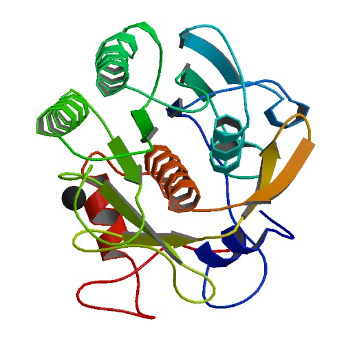
Proteinase K Ribbon Structure ImageSource=RCSB PDB; StructureID=4b5l; DOI=http://dx.doi.org/10.2210/pdb4b5l/pdb;

If you enter any molecular lab asking to borrow some Proteinase K, lab members are likely to answer: “I know we have it. Let me see where it is”. Sometimes the enzyme will be found to have expired. The lab may also have struggled with power outages or freezer malfunctions in the past. But the lab still decides to keep the enzyme. One may rightly ask – why do labs hang on to Proteinase K even when it has been stored under sub-standard conditions?

Continue reading “ProK: An Old ‘Pro’ That is Still In The Game”