Striking Fear into the Heart of Cardiovascular Disease Using Zebrafish and NanoLuc® Luciferase

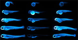
Representative images of ApoB-LP localization in zebrafish across developmental, genetic, pharmacological and dietary manipulations.
Credit: Figure 5.D of The LipoGlo reporter system for sensitive and specific monitoring of atherogenic lipoproteins by James Thierer, Stephen C. Ekker and Steven A. Farber.
Article licensed under Creative Commons Attribution 4.0 International License.

Cardiovascular diseases, or CVDs, are collectively the most notorious gang of cold-blooded killers threatening human lives today. These unforgiving villains, including the likes of coronary heart disease, cerebrovascular disease and pulmonary embolisms, are jointly responsible for more deaths per year than any other source, securing their seat as the number one cause of human mortality on a global scale.

One of the trademarks of most CVDs is the thickening and stiffening of the arteries, a condition known as atherosclerosis. Atherosclerosis is characterized by the accumulation of cholesterol, fats and other substances, which together form plaques in and on the artery walls. These plaques clog or narrow your arteries until they completely block the flow of blood, and can no longer supply sufficient blood to your tissues and organs. Or the plaques can burst, setting off a disastrous chain reaction that begins with a blood clot, and often ends with a heart attack or stroke.

Given the global prevalence and magnitude of this problem, there is a significant and urgent demand for better ways to treat CVDs. In a recent study published in Nature Communications, researchers at the Carnegie Institution for Science, Johns Hopkins University and Mayo Clinic are taking the fight to CVDs through the study of low-density lipoproteins (LDLs), the particles responsible for shuttling bad cholesterol throughout the bloodstream.

Continue reading “Striking Fear into the Heart of Cardiovascular Disease Using Zebrafish and NanoLuc® Luciferase”
I Have My Luciferase Vector, Now What?

Choosing and Optimizing Transfection Methods

Here in Technical Services we often talk with researchers at the beginning of their project about how to carefully design and get started with their experiments. It is exciting when you have selected the Luciferase Reporter Vector(s) that will best suit your needs; you are going to make luminescent cells! But, how do you pick the best way to get the vector into your cells to express the reporter? What transfection reagent/method will work best for your cell type and experiment? Do you want to do transient (short-term) transfections, or are you going to establish a stable cell line?

Continue reading “I Have My Luciferase Vector, Now What?”

Prions Go Slow with ASOs: Experimental Treatment for ALS, Alzheimer’s and Other Prion-like Diseases

In the late-80’s through the 90’s, food and health agencies focused on a mysterious fatal brain disease that infected thousands of cattle. Bovine spongiform encephalitis—or “mad cow disease”—is caused by an infectious protein called a prion. Despite fears that tainted meat would cause the disease to spread to humans, mad cow disease never really made an impact on human health. However, forms of the prion disease such as Creutzfeldt-Jakob disease do affect humans.

In addition to Creutzfeldt-Jakob disease, many neurodegenerative diseases such as Alzheimer’s, Parkinson’s, Huntington’s and amyotrophic lateral sclerosis (ALS or Lou Gehrig’s disease) are now thought to be a result of prion-like activity. There is no cure for these diseases, however, new experimental treatment strategies might help slow the progression of neural degeneration.

prion_bse_histology
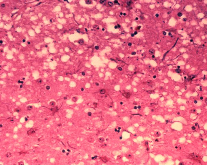
The tell-tale “holes” of prion infection in brain tissue.
Continue reading “Prions Go Slow with ASOs: Experimental Treatment for ALS, Alzheimer’s and Other Prion-like Diseases”

Immunotherapy—Don’t Forget the Microbiome

Bacteria make you sick. The idea that bacteria cause illness has become ingrained in modern society, made evident by every sign requiring employees to wash their hands before leaving a restroom and the frequent food recalls resulting from pathogens like E. coli. But a parallel idea has also taken hold. As microbiome research continues to reveal the important role that bacteria play in human health, we’re starting to see the ways that the microbiota of the human body may be as important as our genes or environment.

The story of how our microbiome affects our health continues to get more complex. For example, researchers are now beginning to understand that the composition of bacteria residing in your body can significantly impact the effects of therapeutic drugs. This is a new factor for optimizing drug response, compared to other considerations such as diet, interaction with other drugs, administration time and comorbidity, which have been understood much longer.

Continue reading “Immunotherapy—Don’t Forget the Microbiome”

Brazilian University Swatting at Leishmaniasis Parasite

The Medicinal Chemistry Center (CQMED), headquartered at Campinas State University in Brazil, recently started a project in partnership with Promega to develop drugs that can be used against Leishmania. This genus of protozoans is the etiological agent of leishmaniasis, transmitted to humans by sandflies.

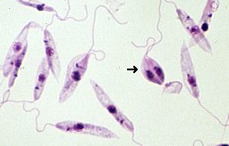
Microscopic image of Leishmania parasite
Microscopic image of Leishmania tropica. Credit: Brian E. Keas at Michigan State University.

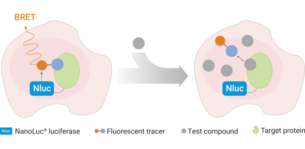
Leishmaniasis is classified as a neglected tropical disease that mainly affects poor communities. Symptoms include large skin sores and an enlarged spleen. The challenge in developing drugs to treat Leishmania is finding appropriate therapeutic targets. These targets are normally proteins whose inhibition leads to death of the parasite. In addition to pharmaceutical company Eurofarma, whose goal is to develop drugs for Leishmania, Promega was chosen to help solve this problem because of our NanoBRET™ Target Engagement (TE) assay*, a well-established technique for measuring protein interactions. In this assay, NanoLuc® luciferase is attached to the protein of interest, and a fluorescent NanoBRET™ tracer molecule is added to the cells. This produces a BRET signal. When a competing ligand is added, it will displace the tracer molecule, enabling quantification of the strength of the interaction compared to the tracer molecule..

A challenge that researchers will face will be ensuring that the NanoBRET™ tracer reaches the inside of the parasite cells; because Leishmania is an intracellular parasite, molecules need to cross the host cell membrane, the membrane of the vacuole containing the parasites, and the membrane of the parasite itself. Another challenge the slow reproduction of Leishmania within macrophages. On top of that is the fact that the parasite’s metabolism varies depending on its biological cycle, meaning that there could be long periods of time during which a drug’s therapeutic target is not expressed in the cell, during which time the drug would have no effect. The ideal target would be expressed at high levels throughout the cell cycle.

The project is being led by Rafael Couñago, a researcher at CQMED, and Promega scientists Matt Robers and Jean-Luc Vaillaud.

*An earlier version of this blog incorrectly said that these experiments are based on the NanoBRET™ assay using HaloTag® protein.

CRISPR/Cas9, NanoBRET and GPCRs: A Bright Future for Drug Discovery

GPCRs

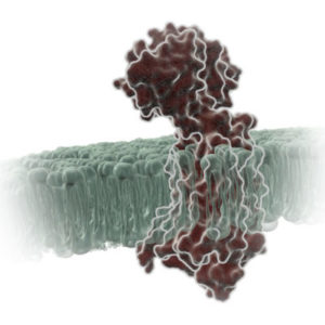
G protein-coupled receptors (GPCRs) are a large family of receptors that traverse the cell membrane seven times. Functionally, GPCRs are extremely diverse, yet they contain highly conserved structural regions. GPCRs respond to a variety of signals, from small molecules to peptides and large proteins. Many GPCRs are involved in disease pathways and, not surprisingly, they present attractive targets for both small-molecule and biologic drugs.

In response to a signal, GPCRs undergo a conformational change, triggering an interaction with a G protein—a specialized protein that binds GDP in its inactive state or GTP when activated. Typically, the GPCR exchanges the G protein-bound GDP molecule for a GTP molecule, causing the activated G protein to dissociate into two subunits that remain anchored to the cell membrane. These subunits relay the signal to various other proteins that interact with or produce second-messenger molecules. Activation of a single G protein can result, ultimately, in the generation of thousands of second messengers.

Given the complexity of GPCR signaling pathways and their importance to human health, a considerable amount of research has been devoted to GPCR interactions, both with specific ligands and G proteins.

Continue reading “CRISPR/Cas9, NanoBRET and GPCRs: A Bright Future for Drug Discovery”

Activating the Inflammasome: A New Tool Brings New Understanding

Innate immunity, the first line of immune defense, uses a system of host pattern recognition receptors (PRRs) to recognize signals of “danger” including invariant pathogen-associated molecular patterns (PAMPs) and damage-associated molecular patterns (DAMPs). These signals in turn recruit and assemble protein complexes called inflammasomes, resulting in the activation of caspase-1, the processing and release of the pro-inflammatory cytokines IL-1ß and IL-18, and the induction of programmed, lytic cell death known as pyroptosis.

Innate immunity and the activity of the inflammasome are critical for successful immunity against a myriad of environmental pathogens. However dysregulation of inflammasome activity is associated with many inflammatory diseases including type 2 diabetes, obesity-induced asthma, and insulin resistance. Recently, aberrant NLRP3 inflammasome activity also has been associated with age-related macular degeneration and Alzheimer disease. Understanding the players and regulators involved in inflammasome activity and regulation may provide additional therapeutic targets for these diseases.

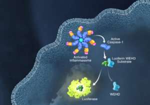
Currently inflammasome activation is monitored using antibody-based techniques such as Western blotting or ELISA’s to detect processed caspase-1 or processed IL-1ß. These techniques are tedious and are only indirect measures of caspase activity. Further, gaining information about kinetics—relating inflammasome assembly, caspase-1 activation and pyroptosis in time—is very difficult using these methods. O’Brien et al. describe a one-step, high-throughput method that enables the direct measurement of caspase-1 activity. The assay can be multiplexed with a fluorescent viability assay, providing information about the timing of cell death and caspase-1 activity from the same sample. Continue reading “Activating the Inflammasome: A New Tool Brings New Understanding”

B Cells, T Cells and Now X Cells?

The cause of type 1 diabetes (T1D) is not well understood. What is known is that in T1D, immune cells attack pancreatic islet cells that produce insulin. In addition, insulin is an autoantigen that activates T cells in diabetic persons.

A new discovery by Ahmed et al. could further T1D understanding. These findings are also setting B and T cell paradigms on their ear.

About B Cells and T Cells

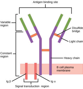
Components of the B cell receptor.
Components of the B-cell receptor.Image by CNX OpenStax. Used with permission under Wikimedia Commons.

B cells (B lymphocytes) are part of the cellular immune response. They act by means of surface receptor molecules that are immunoglobulins. These B cell receptors are created by highly variable gene rearrangements that result in a huge variety of these surface immunoglobulin molecules. The beauty of B cell receptors (BCR) lies in the fact that, through random gene rearrangements comes a such large variety of B cell surface receptors, that any foreign antigen that makes its way into the body is recognized and snagged by a B cell receptor.

B cells then internalize, process and present these antigens to T cells. Continue reading “B Cells, T Cells and Now X Cells?”

Lighting Up GPCR Research with Bioluminescent Tagging

G Protein-Coupled Receptors (GPCRs) are a very large, diverse family of transmembrane receptors in eukaryotes. These receptors detect molecules outside the cell and activate internal signaling pathways by coupling with G proteins. Once a GPCR is activated, β-arrestins translocate to the cell membrane and bind to the occupied receptor, uncoupling it from G proteins and promoting its internalization.

Reporter tags are useful for studying the dynamics of GPCRs and associated proteins, but large tags can disrupt the receptors’ native functioning, and often overexpression of the tagged protein is required to obtain sufficient signal. Here is one example of how researchers have used the small, bright NanoLuc® luciferase to overcome these common challenges and answer questions about GPCRs.

Continue reading “Lighting Up GPCR Research with Bioluminescent Tagging”

Why You Don’t Need to Select a Wavelength for a Luciferase Assay

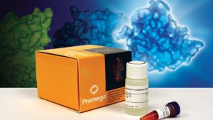
Promega kit depicted; test involves wavelength for a luciferase assay.

It’s a question I’m asked probably once a week. “What wavelength do I select on my luminometer when performing a luciferase assay?” The question is a good and not altogether unexpected one, especially for those new to bioluminescent assays. The answer is that in most cases, you don’t and in fact shouldn’t select a wavelength (the exception to this rule is if you’re measuring light emitted in two simultaneous luciferase reactions). To understand why requires a bit of an explanation of absorbance, fluorescence, and luminescence assays, and the differences among them.

Absorbance, fluorescence, and luminescence assays are all means to quantify something of interest, be that a genetic reporter, cell viability, cytotoxicity, apoptosis, or other markers. In principle, they are all similar. For example, a genetic reporter assay is an indicator of gene expression. The promoter of a gene of interest can be cloned upstream of a reporter such as β-galactosidase, GFP, or firefly luciferase. The amount of each of these reporters that is transcribed into mRNA and translated into protein by the cell is indicative of the endogenous expression of the gene of interest.

Continue reading “Why You Don’t Need to Select a Wavelength for a Luciferase Assay”