The Race to Develop New Therapeutics Against Coronaviruses

Once the purview of virology researchers, the word “coronavirus” is now part of the vernacular in the mainstream media as reports of quarantined cruise ships (1) and makeshift hospitals (2) fill our online news feeds. While there is currently no approved anti-viral treatment for coronavirus infection (3), a team led by researchers from Vanderbilt University recently published work characterizing the anti-CoV activity of a compound, which they now plan to test against 2019-nCoV (4).

Developing New Therapeutics Against Coronaviruses

Coronaviruses (CoVs) are enveloped, single-stranded RNA viruses that exhibit cross-species transmission—the ability to spread quickly from one host (e.g., civet) to another (e.g., human). Scientists classify CoVs into four groups based on the nature of the spikes on their surface: alpha (α), beta (ß), gamma (γ) and delta (δ, 1). Only the alpha- and beta-CoVs can infect humans. Four coronaviruses commonly circulate within human populations: Human CoV 229E (HCoV229E), HCoVNL63, HCoVOC43, and HCoVHKU1. Three other CoVs have emerged as infectious agents, jumping from their normal animal host species to humans: SARS-CoV, MERS-CoV and most recently, 2019-nCoV (5).
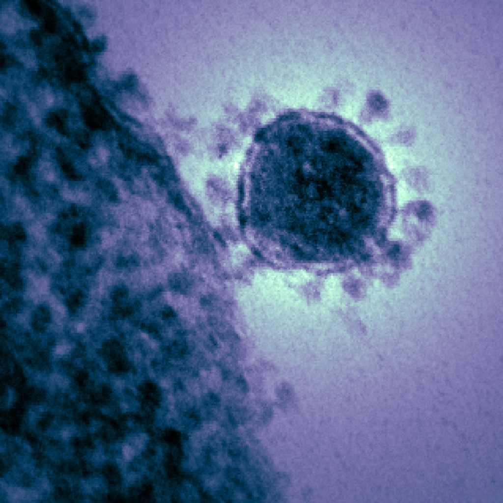
TE micrograph of a single MERS-CoV
Digitally colorized transmission electron micrograph reveals ultrastructural details of a single Middle East respiratory syndrome coronavirus (MERS-CoV) virion. Image credit: National Institute of Allergy and Infectious Diseases

The need for an effective, broad spectrum treatment against HCoVs, has been brought into sharp focus by the recent outbreak of the 2019 Novel Coronavirus (2019-nCoV; 6).

Continue reading “The Race to Develop New Therapeutics Against Coronaviruses”
Targeted Protein Degradation: A Bright Future for Drug Discovery

targeted protein degradation and protacs

Our cells have evolved multiple mechanisms for “taking out the trash”—breaking down and disposing of cellular components that are defective, damaged or no longer required. Within a cell, these processes are balanced by the synthesis of new components, so that DNA, RNA and proteins are constantly undergoing turnover.

Proteins are degraded by two major components of the cellular machinery. The discovery of the lysosome in the mid-1950s provided considerable insight into the first of these degradation mechanisms for extracellular and cytosolic proteins. Over the next several decades, details of a second protein degradation mechanism emerged: the ubiquitin-proteasome system (UPS). Ubiquitin is a small, highly conserved polypeptide that is used to selectively tag proteins for degradation within the cell. Multiple ubiquitin tags are generally attached to a single targeted protein. This ill-fated, ubiquitinated protein is then recognized by the proteasome, a large protein complex with proteolytic activity. Ubiquitination is a multistep process, involving several specialized enzymes. The final step in the process is mediated by a family of ubiquitin ligases, known as E3.

Continue reading “Targeted Protein Degradation: A Bright Future for Drug Discovery”

Designing a Reporter Construct for Analyzing Gene Regulation

Bioluminescent reporter assays are an excellent choice for analyzing gene regulation because they provide higher sensitivity, wider dynamic range and better signal-to-background ratios compared to colorimetric or fluorescent assays. In a typical genetic reporter assay, cells are transfected with a vector that contains the sequence of interest cloned upstream of a reporter gene, and the reporter activity is used to determine how the target sequence influences gene expression under experimental conditions. A second control reporter encoded on the same or a different plasmid is an essential internal control. The secondary reporter is used to normalize the data and compensate for variability caused by differences in cell number, lysis efficiency, cell viability, transfection efficiency, temperature, and measurement time. 

Basic Introduction to the Strategy of Reporter Gene Assays

For genetic reporter assays, using a secondary control vector with a weak promoter like PGK or TK to ensures that the control does not interfere with activation of your primary reporter vector. Transfection of high amounts of the control plasmid or putting the control reporter under control of a strong promoter like CMV or SV40 often leads to transcriptional squelching or other interference with the experimental promoter (i.e., trans effects). Reporter assays can also be used to quantitatively evaluate microRNA activity by inserting miRNA target sites downstream or 3´ of the reporter gene. For example, the pmirGLO Dual-Luciferase miRNA Target Expression Vector is based on dual-luciferase technology, with firefly luciferase as the primary reporter to monitor mRNA regulation and Renilla luciferase as a control reporter for normalization.

Here in Technical Services we often talk with researchers who are just starting their project and looking for advice on designing their genetic reporter vector. They have questions like:

  • How much of the upstream promoter region should be included in the vector?
  • How many copies of a response element will be needed to provide a good response?
  • Does the location of the element or surrounding sequence alter gene regulation?
Continue reading “Designing a Reporter Construct for Analyzing Gene Regulation”

Infographic: Assays for Measuring Insulin Activity

We rely on insulin supplied by our pancreas at the right dose and at the right time to control our blood glucose levels and energy storage. Insulin works by regulating the energy usage of various cell types in the body. When this process goes awry, it can cause diabetes.

There are two types of diabetes, defined by how insulin is dysregulated. In Type 1 Diabetes (T1D), the pancreas produces too little insulin. Patients need to give themselves insulin in order to respond to glucose in the diet. In Type 2 Diabetes (T2D), patients do not respond well to the insulin produced in their body. Therefore, they need to give themselves more to avoid hyperglycemia (high blood glucose).

Continue reading “Infographic: Assays for Measuring Insulin Activity”

NanoLuc: Tiny Tag with a Big Impact

Synthetic biology—genetically engineering an organism to do or make something useful—is the central goal of the iGEM competition each year. After teams conquer the challenge of cloning their gene, the next hurdle is demonstrating that the engineered gene is expressing the desired protein (and possibly quantifying the level of expression), which they may do using a reporter gene.

Reporters can also play a more significant role in iGEM projects when teams design their organism with reporter genes to detect and signal the presence of specific molecules, like environmental toxins or biomarkers. Three of the iGEM teams Promega sponsored this year opted to incorporate some version of NanoLuc® Luciferase into their projects.

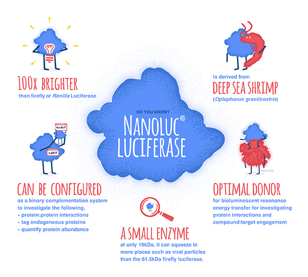
NanoLuc® luciferase is a small monomeric enzyme (19.1kDa, 171 amino acids) based on the luciferase from the deep sea shrimp Oplophorus gracilirostris. This engineered enzyme uses a novel substrate, furimazine, to produce high-intensity, glow-type luminescence in an ATP-independent reaction. Unlike other molecules for tagging and detecting proteins, NanoLuc® luciferase is less likely to interfere with enzyme activity and affect protein production due to its small size.

NanoLuc® Luciferase has also been engineered into a structural complementation reporter system, NanoBiT® Luciferase, that contains a Large subunit (LgBiT) and two small subunit options: low affinity SmBiT and high affinity HiBiT. Together, these NanoLuc® technologies provide a bioluminescent toolbox that was used by the iGEM teams to address a diverse set of biological challenges.

Here is an overview of each team’s project and how they incorporated NanoLuc® technology.

Continue reading “NanoLuc: Tiny Tag with a Big Impact”

Out-FOXOing High-Stage Neuroblastoma

Fluorescence microscopy of neuroblastoma cells.

In recent years, scientists have been hot on the trail of transcription factor FOXO3, tracing its involvement in various tumor-centric activities comprising the many trademarks of cancer, from drug resistance to metastasis to tumor angiogenesis.

FOXO3 is a member of the O sub-class of the forkhead box family of transcription factors. The forkhead box (FOX) family is characterized by a fork head DNA-binding domain (DBD), comprised of around 100 amino acids. They have also proven themselves to be a family of many hats, functioning in diverse roles ranging from metabolism, immunology, cell-cycle control, development, as well as cancer (1). The forkhead box O (FOXO) sub-class alone has demonstrated involvement in a variety of cellular outcomes, from drug resistance and longevity to apoptosis induction.

Due to its pro-apoptotic and anti-proliferative proclivity, FOXO3 has been previously identified as a tumor suppressor gene. However, more and more studies have begun to flip the narrative on FOXO3, portraying it more as a devoted henchman, due to its roles in drug and radiotherapy resistance, cell-cycle arrest and long-term maintenance of leukemia-initiating stem cells in a variety of cancer types, including breast cancer, pancreatic cancer, glioblastoma, and both acute and chronic myeloid leukemia.

Continue reading “Out-FOXOing High-Stage Neuroblastoma”

MSI Testing of Tumor Cells for Better Tailored Treatment

3D artistic rendering of mismatch repair. Microsatellite instability (MSI)  which can result from defects in mismatch repair is a biomarker for some cancers

There are as many different cancers as there are people with cancer. Unlike infectious diseases, which are caused by pathogens that are foreign to our bodies (bacteria, viruses, parasites), cancer cells arise from our body—our own cells gone rogue. Because cancer is a dysfunction of a person’s normal cells, every cancer reflects the genetic differences that mark us as individuals. Add to that environmental influences like diet, tobacco use, the microbiome and even occupation, and the likelihood of finding a “single” pharmaceutical cure for cancer becomes virtually impossible.

But, while looking for a single cure for all cancers may not be a fruitful activity, defining a best practice for understanding the genetic and protein biomarkers of individual tumors is proving worthwhile.

Continue reading “MSI Testing of Tumor Cells for Better Tailored Treatment”

Discussing the Future of Gene Editing at CRISPRcon Midwest

crisprcon_banner
Walking in to the first session at CRISPRcon Midwest.

Last week, a diverse group of stakeholders attended CRISPRcon Midwest, hosted by the Keystone Policy Center and the University of Wisconsin–Madison. The goal of the day-long conference was to emphasize the importance and value of gene editing technology, and how it must be communicated deliberately between scientists, the public, policymakers, and other stakeholders.

Julie Shapiro, Senior Policy Director of Keystone Policy Center, acted as Emcee for the event. Given the diverse group of attendees, she mentioned in her opening remarks that the event organizers were “seeking conversation, not consensus” and emphasized the “power of respectful dialogue.” A slide overhead showcased the ground rules for the day, which included statements such as “dare to listen, dare to share, and dare to disagree.”

crisprcon_poll_word_cloud
Word cloud generated from live polling results at CRISPRcon Midwest.

CRISPRcon aimed to included voices beyond those represented by keynote speakers and panelists, so they incorporated live polling through an online app to keep the audience engaged and an active participant in the conversations throughout the day. From the opening remarks, it was clear that this conference would not just deliver on its promise of thoughtful conversation about the science, but build further understanding about the societal impacts of a rapidly advancing technology.

Continue reading “Discussing the Future of Gene Editing at CRISPRcon Midwest”

All You Need is a Tether: Improving Repair Efficiency for CRISPR-Cas9 Gene Editing

Ribonucleoprotein complex with Cas9, guide RNA and donor ssDNA. Copyright Promega Corporation.

With the advent of genome editing using CRISPR-Cas9, researchers have been excited by the possibilities of precisely placed edits in cellular DNA. Any double-stranded break in DNA, like that induced by CRISPR-Cas9, is repaired by one of two pathways: Non-homologous end joining (NHEJ) or homology-directed repair (HDR). Using the NHEJ pathway results in short insertions or deletions (indels) at the break site, so the HDR pathway is preferred. However, the low efficiency of HDR recombination to insert exogenous sequences into the genome hampers its use. There have been many attempts at boosting HDR frequency, but the methods compromise cell growth and behave differently when used with various cell types and gene targets. The strategy employed by the authors of an article in Communications Biology tethered the DNA donor template to Cas9 complexed with the ribonucleoprotein and guide RNA, increasing the local concentration of the donor template at the break site and enhancing homology-directed repair. Continue reading “All You Need is a Tether: Improving Repair Efficiency for CRISPR-Cas9 Gene Editing”

NLRP3: The New Hope for Treating Chronic Inflammatory Diseases

Inflammasome - inflammatory diseases caused by NLRP3

Our innate immune system was meant to do good. Up until a century ago, most humans died from infectious diseases like diarrhea, tuberculosis and meningitis. Over millions of years, our immune system has evolved to fight these life-threatening infections from pathogens. As a result, we have developed a highly efficient response to these tiny invaders. But it seems that our immune system may be turning against us.

Continue reading “NLRP3: The New Hope for Treating Chronic Inflammatory Diseases”