Seeing is Believing: How NanoLuc® Luciferase Illuminates Virus Infections

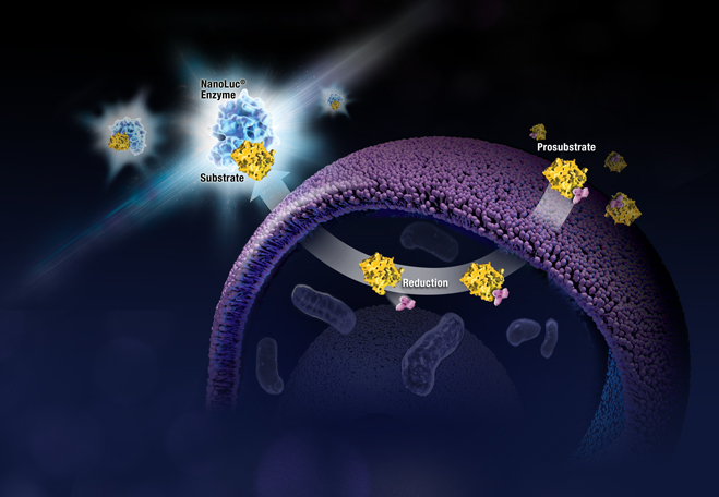
Artists interpretation of in vivo imaging of viral infections in mice using NanoLuc luciferase.

Wearing blue surgical gowns and white respirator hoods, research scientist Pradeep Uchil and post-doctoral fellow Irfan Ullah carry an anesthetized mouse to the lab’s imaging unit. Two days ago, the mouse was infected with a SARS-CoV-2 virus engineered to produce a bioluminescent protein. After an injection of a bioluminescence substrate, a blue glow starts to emanate from within the mouse’s nasal cavity and chest, visible to the imaging unit’s camera and Uchil’s eyes.

“We were never able to see this kind of signal with retrovirus infections.” Uchil is a research scientist at the Yale School of Medicine whose work focuses on the in vivo imaging of retroviral infections. Normally, the mouse would have to be sacrificed and “opened up” for viral bioluminescent signals from internal tissues to be imaged directly.

Continue reading “Seeing is Believing: How NanoLuc® Luciferase Illuminates Virus Infections”

3D Cell Culture Models: Challenges for Cell-Based Assays

3D Cell Culture Spheroid

In 3D cell culture models, cells are grown under conditions that allow the formation of multicellular spheroids or microtissues. Instead of growing in a monolayer on a plate surface, cells in 3D culture grow within a support matrix that allows them to interact with each other, forming cell:cell connections and creating an environment that mimics the situation in the body more closely than traditional 2D systems. Although 3D cultures are designed to offer a more physiologically accurate environment, the added complexity of that environment can also present challenges to experimental design when performing cell-based assays. For example, it can be a challenge for assay reagents to penetrate to the center of larger microtissues and for lytic assays to disrupt all cells within the 3D system.

Earlier this week Terry Riss, a Senior Product Specialist at Promega, presented a Webinar on the challenges of performing cell-based assays on microtissues in 3D cell culture. During the Webinar, Terry gave an overview of the different methods available for 3D cell culture, providing a description of the advantages of each. He then discussed considerations for designing and optimizing cell-based  assays for use in 3D culture systems, providing several  recommendations to keep in mind when performing cell viability assays on larger microtissue samples.

Continue reading “3D Cell Culture Models: Challenges for Cell-Based Assays”

Permeability Possibilities with the New NanoClick Assay

Peptide Predicament

For decades now, peptides have been a molecule of interest for drug discovery research. Peptides offer a unique opportunity for therapeutic intervention that closely mimics natural pathways, as many physiological functions utilize peptides as intrinsic signaling molecules. Macrocyclic peptides, in particular, have recently proven to be promising candidates for targeting intracellular protein–protein interactions (PPIs), an attractive but hard-to-reach therapeutic target for conventional small molecule and biological drugs.  

As with any opportunity, there are also challenges that accompany the peptide therapeutic development. Peptide ligands typically have poor membrane permeability, so thus far the majority of peptide therapeutics predominantly target extracellular proteins and receptors. There are also multiple mechanisms for cellular uptake of peptides, including both energy-dependent routes like endocytosis, and energy-independent, like passive diffusion or membrane translocation. Multiple mechanisms of cellular uptake paired with poor permeability makes engineering enough membrane permeability into peptides in order to advance them through drug discovery pipelines extremely difficult.  

There are other factors to consider in developing peptide therapeutics, such as solubility, protein/lipid binding and stability, which can also have an affect on the overall cytosolic concentration and, ultimately impact the ability of the peptide to effectively engage its desired intracellular targets.

With so many challenging factors, the ability to have a predictive, high-throughput assay to assess cell permeability, independent of the mechanism(s) of entry, would be a critical and invaluable tool to support peptide drug discovery research.

In a recent study published in ACS Chemical Biology, researchers sought to develop such a tool, and demonstrated a new application for Promega NanoBRET™ technology: the NanoClick assay.

Continue reading “Permeability Possibilities with the New NanoClick Assay”

The NLRP3 Inflammasome: Flipping the Switch

It’s been just over 10 years since the world lost a pioneering immunologist and biochemist, Dr. Jürg Tschopp. He died tragically during a hiking trip in the Swiss Alps on March 22, 2011. A host of academic journals, including Science, Nature and Cell, paid tribute to Dr. Tschopp with obituaries that highlighted his many accomplishments in the fields of apoptosis and immunology.

In 2002, a team led by Dr. Tschopp at the University of Lausanne, Switzerland, was studying the role of the proinflammatory cytokine interleukin 1 beta (IL-1β). This cytokine is produced in the cytoplasm as an inactive precursor (pro-IL-1β). It is cleaved by caspase-1 to the active form, but the exact process by which caspase-1 itself is activated was unknown at the time. Several members of the caspase family contain a conserved region known as the caspase recruitment domain or CARD, and it was proposed that this domain was essential to caspase activation.

Based on similarity to another protein containing an N-terminal CARD motif (Apaf-1) that is involved in activation of caspase-9, the researchers examined the roles of a family of proteins known as NALP1, NALP2 and NALP3 (1). In particular, they were interested in NALP1, which is involved in the immune response. Unlike Apaf-1, NALP1 contains a CARD motif at the C terminus, while the N terminus contains a related motif known as a pyrin-like domain (PYD). The research team had previously showed that the PYD region of NALP1 interacted with an adapter protein known as PYCARD or ASC, which also contains an N-terminal PYD and C-terminal CARD.

The results of the team’s in vitro binding, activation and immunodetection studies showed that a multi-unit protein complex is responsible for caspase activation, and they called this complex the “inflammasome” (1). It is composed of caspase-1, caspase-5, PYCARD/ASC and NALP1.

Continue reading “The NLRP3 Inflammasome: Flipping the Switch”

Immune Checkpoint Bioassays Strengthen Cancer Research in the Development of New Therapies

This post was written by guest blogger, Nicole Werner, Product Management Support at Promega GmbH.

“You have cancer.” – a statement that fundamentally changes life in a second. After the first shock, the insight often arises: “If only I had stopped smoking sooner!”

Lung cancer, while not the leading cause of death worldwide, is the leading preventable cause of death in developed countries. According to the WHO, eight million people die each year as a result of smoking, including one million as a result of passive smoking [1]. Currently, 80% of those affected die within the next 13 months after diagnosis [1]. New therapeutic approaches, such as treatment with immune checkpoint inhibitors, bring hope.

Promega supports research in this area with the high-precision tools needed to develop this new form of therapy.

Artistic 3D rendering  of Immune checkpoint signaling. Immune checkpoint bioassays enable researcher to characterize  therapeutic antibodies trageting these pathways.
Continue reading “Immune Checkpoint Bioassays Strengthen Cancer Research in the Development of New Therapies”

Overcoming Challenges to Detect Apoptosis in 3D Cell Structures

This blog is written by guest author, Maggie Bach, Sr. Product Manager, Promega Corporation.

Researchers are increasingly relying on cells grown in three-dimensional (3D) structures to help answer their research questions. Monolayer, or 2D cell culture, was the go-to cell culture method for the past century. Now, the need to better represent in vivo conditions is driving the adoption of 3D cell culture models. Cells grown in 3D structures better mimic tissue-like structures, better exhibit differentiated cellular functions, and better predict in vivo responses to drug treatment.

Switching to 3D cell culture models comes with challenges. Methods to interrogate these models need to be adaptable and reliable for the many types of 3D models. Some of the most popular 3D models include spheroids grown in ultra-low attachment plates, and cells grown in an extracellular matrix, such as Matrigel® from Corning. Even more complex models include medium flow over the cells in microfluidic or organ-on-a-chip devices. Will an assay originally developed for cells grown in monolayer perform consistently with various 3D models? How is measuring a cellular marker different when cells are grown in 3D models compared to monolayer growth?

Close up of cells in 3D culture apparatus. 3D Cell Structures Provide Challenges for Measuring Markers of Cellular Activitiy
3D Cell Structures Provide Challenges for Measuring Markers of Cellular Activitiy
Continue reading “Overcoming Challenges to Detect Apoptosis in 3D Cell Structures”

Bringing Cutting Edge Technologies to Academic Researchers Through the Academic Access Program

This post was written by guest blogger Iain Ronald, Director Academic/Government Market Segment at Promega.

My back story is similar to most of you reading this blog, high school education, undergraduate degree then onto a postgraduate degree. However, over 25 years ago during my undergraduate study, I was fortunate enough to work in the lab of Professor Ray Waters studying DNA damage in S. cerevisiae as a model organism and at the time PCR was cutting-edge technology and the PCR license was in full effect. However, there was one company that was fighting the good fight to democratize PCR for the good of the scientific community, Promega.

I became enamored with Promega then, and the next steps in my career were taken with a view to working at this company who, for all intents and purposes, seemed to really care about the progression of science beyond self-aggrandizement.

Now that I am working at Promega in a position where I can bring benefit to our academic community, I have pondered what I can do to equal the disruptive attitude I observed in this company all those years ago when they were fighting the then “big tech” for the enablement of the scientific community. 

Reporter bioassays are one of hte many offerings of the academic access program.
Continue reading “Bringing Cutting Edge Technologies to Academic Researchers Through the Academic Access Program”

How Promega Helped Our Lab Scale Up Drug Discovery for Bloodborne Pathogens

This blog was written by Sebastien Smick, Research Technician in Dr. Jacquin Niles’ laboratory at Massachusetts Institute of Technology (MIT)

Our lab is heavily focused on the basic biology and drug discovery of the human bloodborne pathogen Plasmodium falciparum, which causes malaria. We use the CRISPR/Cas9 system, paired with a TetR protein fused to a native translational repressor alongside a Renilla luciferase reporter gene, to conditionally knock down genes of interest to create modified parasites. We can then test all kinds of compounds as potential drug scaffolds against these gene-edited parasites. Our most recent endeavor, one made possible by Promega, is a medium-low throughput robotic screening pipeline which compares conditionally-activated or-repressed parasites against our dose-response drug libraries in a 384-well format. This process has been developed over the past few years and is a major upgrade for our lab in terms of data production. Our researchers are working very hard to generate new modified parasites to test. Our robots and plate readers rarely get a day’s rest!

Continue reading “How Promega Helped Our Lab Scale Up Drug Discovery for Bloodborne Pathogens”

Toxicity Studies in Organoid Models: Developing an Alternative to Animal Testing

Alternatives to animal testing have long been explored when it comes to studying the safety of various chemical compounds for use in food, medicine and cosmetics. With the advent of three-dimensional (3D) cell culture to create organoids, more relevant human organoid models are being explored as one way to provide safe and effective compound testing while minimizing the need for testing in animals. The international project Physiologically Anchored Tools for Realistic nanOmateriaL hazard aSsessment (PATROLS) led by the Swansea University Medical School aims to establish a battery of innovative, next-generation safety testing tools that can more accurately predict the effects of engineered nanomaterial (ENM) exposure in humans and the environment.

One project being researched by Samantha Llewellyn, a research assistant at Swansea University, is developing predictive physiologically relevant 3D liver models for ENM safety assessment. By having a model to evaluate realistic ENM exposures, a researcher can study liver function, hepatic metabolism and microtissue cell viability after acute (24 hours) or prolonged (several days) exposure. A microtissue model for assessing ENM hepatotoxicity needs to mimic primary hepatocytes and be amenable to assays used to test cell viability and metabolism.

The right tools for testing this 3D liver model include the bioluminescent-based CellTiter-Glo® 3D Viability and P450-Glo® Assays. When creating organoids, having reagents that can penetrate to the center of the dense and complex 3D liver spheroids is important so that the cell viability readout encompasses the entire microtissue. The CellTiter-Glo® 3D Viability Assay accomplishes this task, providing accurate assessment of 3D tissue cell health. Measuring cytochrome P450 (CYP450) activity is necessary for studying liver function. The P450-Glo® Assays have the flexibility to assess CYP450 activity while preserving the liver spheroids; thus, researchers can gather more data from a single experiment.

The importance of Samantha Llewellyn’s research as part of PATROLs is establishing a 3D liver model that could evaluate realistic ENM exposures and reduce the need for animal testing. Bioluminescent assays for assessing cell health and liver enzyme function are necessary to reach this goal.

luciferse technologies are allowing researchers to develop predictive 3D organoid models for ERM testing

To learn more about the last 30 years of bioluminescent innovations and the discoveries they’ve enabled, please visit our 30th anniversary celebration page.

Related Posts

Understanding Inflammation: A Faster, Easier Way to Detect Cytokines in Cells

Inflammation, a process that was meant to defend our body from infection, has been found to contribute to a wide range of diseases, such as chronic inflammation, neurodegenerative disorders—and more recently, COVID-19. The development of new tools and methods to measure inflammation is crucial to help researchers understand these diseases.

This diagram shows how the Lumit™ Immuno assay can be used to detect cytokines.

Cytokines—small signaling molecules that regulate inflammation and immunity—have recently become the focus of inflammation research due to their role in causing severe COVID-19 symptoms. In these severe cases, the patient’s immune system responds to the infection with uncontrolled cytokine release and immune cell activation, called the “cytokine storm”. Although the cytokine storm can be treated using established drugs, more research is needed to understand what causes this severe immune response and why only some patients develop it.

Continue reading “Understanding Inflammation: A Faster, Easier Way to Detect Cytokines in Cells”