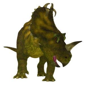
Did dinosaurs get cancer? That isn’t an easy question to answer. Finding and diagnosing cancer in dinosaur fossils has proven difficult. Any soft tissue, the typical location of tumors, has degraded over the millennia. Fossilized bones millions of years old are subject to wear and tear, making it hard to distinguish bone damage from possible pathology. By using the knowledge and expertise gained from diagnosing cancer in humans, a team reported in The Lancet Oncology that they found the first known case of osteosarcoma in a lower leg bone from a horned dinosaur found in southern Alberta, Canada.
This case of bone cancer discovered in a specimen of Centrosaurus apertus found in the Canadian Dinosaur Park Formation was confirmed by examining the bone surface along with radiographic and histological analysis. The 77–75.5-million-year-old case was compared to both a normal C. apertus fibula from the Oldman formation also in southern Alberta, Canada, as well as a human fibula with an osteosarcoma.
Specimen Examination
Starting with the gross pathological exam, both the human and possible C. apertus fibula show an abnormal mass on the bone on the proximal end. The dinosaur fibula was not intact, missing part of the upper bone. This suggested that the changes could be the result of a fracture. In the case of the human osteosarcoma, x-rays, magnetic resonance imaging (MRI) and computed tomography (CT) analysis showed a large mass surrounding the bone. In addition, the cortex (the outside of the bone) had been destroyed, with islands of bone within the soft tumor tissue. Microscopic examination of the human tumor showed the intramedullary cavity of the bone had been partially replaced by immature bone cells as well as trapped remnants of mature bone cells.
For the C. apertus bone, nearly half of the bone was taken up by the probable tumor mass. High-resolution x-ray CT scan showed destruction of the cortex and changes to medullary cavity. Like that seen with the human osteosarcoma, researchers observed a triangular signature where the periosteum (a layer of vascular connective tissue that envelops bones) was pushed up as a result of the tumor. In addition, there seemed to be abnormalities in new bone formation in the pathological lesion. Spanning the length of the bone, four histological sections of both normal and possible cancerous C. apertus fibula were taken for microscopic analysis. While the unaffected end of the C. apertus fibula looked the same as the normal bone, the morphological changes showed how the tumor intruded into the cortex of the bone.
Diagnosis
By using the same techniques to analyze and diagnose human tumors, the multidisciplinary team was confident in stating that the lesion on this C. apertus fibula was osteosarcoma and not an alternate diagnosis of a fracture, bone infection, noncancerous overgrowth of bone (osteochondroma) or non-ossifying fibroma. While not the oldest cancer diagnosis, this approach highlights that closer examination of unexpected lesions on fossils means researchers can diagnose what may have happened to an animal, even millions of years ago—even cancer in dinosaurs.
Reference
Ekhtiari, S. et al. (2020) First case of osteosarcoma in a dinosaur: A multimodal diagnosis. Lancet Oncol. 21, 1021–2.
Related Posts
Sara Klink
Latest posts by Sara Klink (see all)
- A One-Two Punch to Knock Out HIV - September 28, 2021
- Toxicity Studies in Organoid Models: Developing an Alternative to Animal Testing - June 10, 2021
- Herd Immunity: What the Flock Are You Talking About? - May 10, 2021
