All You Need is Pla (for Pneumonic Plague)

Yersinia pestis. By Mrs Robinson at bg.wikipedia (Transferred from bg.wikipedia) [Public domain], from Wikimedia Commons.
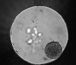
Writing about Yersinia pestis or the Black Death, has earned me a reputation among Promega Connections bloggers. I am interested in what researchers have been able to piece together about the causative agent of ancient plagues, what modern research shows about how Y. pestis spreads in the body and the continuing reservoirs in modern times, resulting in publication of eight blog posts on the subject. Understanding Y. pestis bacterium is of continuing interest to researchers. How did Yersinia pestis evolve from the humble Yersinia pseudotuberculosis, a pathogen that causes gastrointestinal distress, into a virulent pneumonic plague that is a global killer? One strategy for answering this question is to look at the genomic tree of Y. pestis and trace which strains had what characteristics. In a recent Nature Communications article, Zimbler et al. explored the role of the plasmid pPCP1 in Y. pestis evolution and the signature protease Pla it expresses. Continue reading “All You Need is Pla (for Pneumonic Plague)”

Developing a Model System to Test Ketamine Toxicity

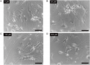
Figure 2. Ketamine induced morphological changes in neurons derived from iPSCs. Cells were treated with 0μM (Panel A), 20μM (Panel B), 100μM (Panel C) or 500μM (Panel D) ketamine for 24 hours. doi:10.1371/journal.pone.0128445.g002
Ketamine induced morphological changes in neurons derived from iPSCs.
Cells were treated with 0μM (Panel A), 20μM (Panel B), 100μM (Panel C) or 500μM (Panel D) ketamine for 24 hours. Scale bar = 50μm. From Ito, H., Uchida, T. and Makita, K. (2015) Ketamine causes mitochondrial dysfunction in human induced pluripotent stem cell-derived neurons. PLOS ONE 10, e0128445.
doi:10.1371/journal.pone.0128445.g002
When I consider that major surgery was performed long before anesthetics were developed, I am grateful to be alive in the anesthesia era. Just the thought of being subjected to various cutting and retracting instruments without general anesthesia calls to mind a phrase: The cure is worse than the disease. Despite the advantages of unconsciousness during surgery, anesthesia can have side effects. Studies in neonatal nonhuman primates have demonstrated that the anesthetic ketamine has toxic effects. However, the differences between humans and nonhuman primates mean the outcome in one species is not the same in another. In an article recently published in PLOS ONE, scientists were interested in creating an experimental model of developing human neurons and using the model to better understand the toxic effects of ketamine on human cells. Continue reading “Developing a Model System to Test Ketamine Toxicity”

Cancer Detection on a Chip?

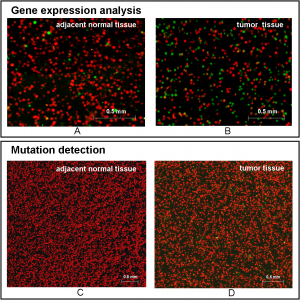
Figure 5 shows typical scanned images of bead-array for analyzing adjacent normal tissue and tumor tissue. Huang et al. (2015) Digital Detection of Multiple Minority Mutants and Expression Levels of Multiple Colorectal Cancer-Related Genes Using Digital-PCR Coupled with Bead-Array. PLOS ONE 10(4):e0123420. doi:10.1371/journal.pone.0123420.g005
The ideal cancer detection method would involve giving a sample of blood or tissue and using DNA or RNA analysis to determine if there were any gene sequence or gene expression changes that are known hallmarks of cancer. Unfortunately, most current screening methods used are not so precise and in some cases are invasive. However useful tests for colon cancer may be, many people do not subject themselves to the standard colonoscopy. What if there was an easier, noninvasive method that could be used to screen for cancer and detect changes at the early, easily treatable stages of cancer? A recent article in PLOS ONE describes just such a mutation detection method for colorectal cancer using purified nucleic acid with a method that involves emulsion PCR, bead arrays and fluorescent probes. Continue reading “Cancer Detection on a Chip?”

Cloning Tips for Restriction Enzyme-Digested Vectors and Inserts
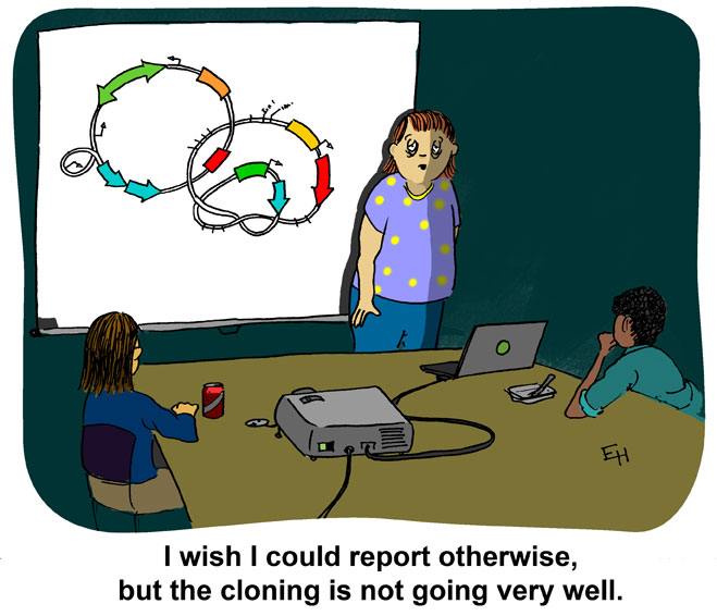
Cartoon created and owned by Ed Himelblau
While T-vector cloning is commonly used for PCR-amplified inserts, restriction enzymes still have their uses. For example, you can ensure directional cloning if you digest a vector with the same two enzymes like BamHI and EcoRI that are used to digest your insert. This way, the insert can only be cloned in one direction. However, there are other cloning techniques that can be used to modify the end of vectors and inserts after restriction enzyme digestion and prior to ligation. Continue reading “Cloning Tips for Restriction Enzyme-Digested Vectors and Inserts”

Could This be the Next Generation Ebola Virus Vaccine?

Ebola virus has received a lot of press in the last year due to the extended epidemic outbreak in Africa. Ebola is part of the family of Filioviruses (filamentous virus) and causes hemorrhagic fever that leads to internal bleeding and loss of bodily fluids. As the epidemic in Africa has illustrated so starkly, once the virus infects a large enough population, the human suffering it causes is devastating to individuals and communities. Because no treatment other than palliative fluid support is available to those infected by Ebola virus, virologists have focused attention on potential therapeutics and vaccines. The vaccine strategies now in clinical trials are based on a single Ebola virus glycoprotein, GP, and involve a DNA-based vaccine or innoculation with an Ebola protein expressed from a viral vector. How effective and safe this approach may be for protection from Ebola virus infection is currently under investigation.

Based on the history of effective vaccines, Marzi et al. was interested in testing a whole-virus vaccine for Ebola (EBOV). A whole-virus-based vaccine like smallpox or measles uses an attenuated or inactivated virus. The advantage of this method is that all the proteins as well as the nucleic acid are available for immunological reaction, offering broader-based protection than a single protein. In the recently published Science report from Marzi et al., a replication-incompetent Ebola virus was used as the basis for a whole-virus vaccine that was tested for its efficacy in nonhuman primates.

Continue reading “Could This be the Next Generation Ebola Virus Vaccine?”

Uncovering Protein Autoinhibition Using NanoBRET™ Technology

13305818-protein ligand

In a study published in Proceedings of the National Academy of Sciences USA article, Wang et al. used the principle of the Promega NanoBRET™ assay to understand how ERK1/2 phosphorylation of Rabin8, a guanine nucleotide exchange factor, influenced its configuration and subsequent activation of Rab8, a protein that regulates exocytosis.
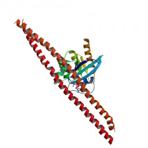
Crystal structure of GDP-boudn Rab8:Rabin8 ImageSource=RCSB PDB; StructureID=4lhy; DOI=http://dx.doi.org/10.2210/pdb4lhy/pdb;

Rab8 is a member of the Rab family of small GTPases and an important regulator of membrane trafficking from the trans Golgi network and recycling endosomes to the plasma membrane. Wang et al. were interested in learning how the guanine nucleotide exchange factor (GEF) Rabin8, a known activator of Rab8, was itself activated to better understand how Rab8 and exocytosis were regulated in the cell. First, they confirmed if the consensus extracellular-signal-regulated kinases ERK1/2 phosphorylation motif uncovered in Rabin8 resulted in phosphorylation of Rabin8. Both in vitro analysis and cell-based assays confirmed that ERK1/2 phosphorylated Rabin8. Next, the GEF activity of Rabin8 was assessed to determine if ERK1/2 phosphorylation activated the GEF. Researchers confirmed activation of Rabin8 GEF in vitro.

Continue reading “Uncovering Protein Autoinhibition Using NanoBRET™ Technology”

Tracking the Beginning of a Pathogenic Bacterial Infection
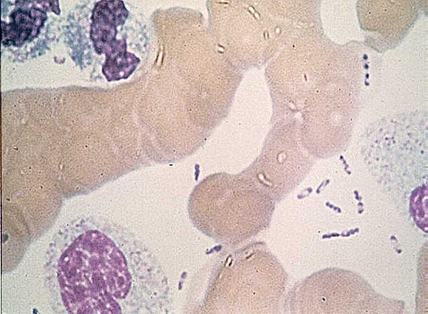
Yersinia pestis by U.S. Center for Disease Control [Public domain], via Wikimedia Commons.
Understanding the course of a pathogenic infection involves not only understanding what ultimately kills the host or how the bacterium or virus enters the body but also how it establishes itself in the host organism. What is the receptor that allows a virus to enter the cell? Which cells does a bacterium first target or how does it evade an immune response? While other studies of bacteria like Yersina pestis have looked at imaging the bacterial burden in model mice, questions remain about how this bacterium gets from the skin after an infected flea bites to the draining lymph nodes, where the bacteria replicate and enter the bloodstream and infection becomes fatal. A recent PLOS Pathogens article examined how the nonmotile Y. pestis disseminated itself starting from a tiny innoculation mimicking a flea bite on a mouse ear and following pathogen interaction with the host from skin to lymph node. Continue reading “Tracking the Beginning of a Pathogenic Bacterial Infection”

General Considerations for Transfection

Many studies, from reporter assays to protein localization to BRET and FRET, require successful transfection first. Yet, transfection can be tricky and difficult. There are many considerations when planning transfection of your cells including reagent selection, stable or transient experiment, type of molecule and endpoint assay used. Here we discuss these considerations to help you plan a successful transfection scheme for your experimental system. Continue reading “General Considerations for Transfection”

Using Laser Treatment to Eliminate Blood-Borne Pathogens

One unit of fresh frozen plasma By DiverDave (Own work) [CC BY-SA 3.0 (http://creativecommons.org/licenses/by-sa/3.0) or GFDL (http://www.gnu.org/copyleft/fdl.html)], via Wikimedia Commons
Keeping our blood supply safe is increasingly difficult in the face of newly discovered pathogens that can be transmitted via blood. The tests developed to ensure the blood is free of pathogens like HIV and HBV are based on known pathogens and available screening tests. What about an emerging virus? How can we ensure our blood is safe in the face of the virus we know nothing about? The PLOS ONE article by Tsen et al. explored how a nonchemical treatment using ultrashort pulsed lasers might be used to eliminate viruses from blood plasma.

The authors used ultrashort pulsed (USP) lasers in their research as this treatment is known to inactivate a spectrum of bacteria and viruses including nonenveloped viruses, a class of virus that resists inactivation. Furthermore, the laser treatment is nonionizing and does not modify proteins covalently, meaning that proteins present in blood are likely to remain active even after exposure to USP lasers. The viruses that were tested for inactivation by USP laser in human plasma were an enveloped RNA virus human immunodeficiency virus (HIV), nonenveloped RNA virus hepatitis A virus (HAV) and enveloped DNA virus murine cytomegalovirus (MCMV). Continue reading “Using Laser Treatment to Eliminate Blood-Borne Pathogens”