Real-Time (quantitative) qPCR for Quantitating Library Prep before NGS

Real-Time (or quantitative, qPCR) monitors PCR amplification as it happens and allows you to measure starting material in your reaction.

This the last in a series of four blogs about Quantitation for NGS is written by guest blogger Adam Blatter, Product Specialist in Integrated Solutions at Promega.

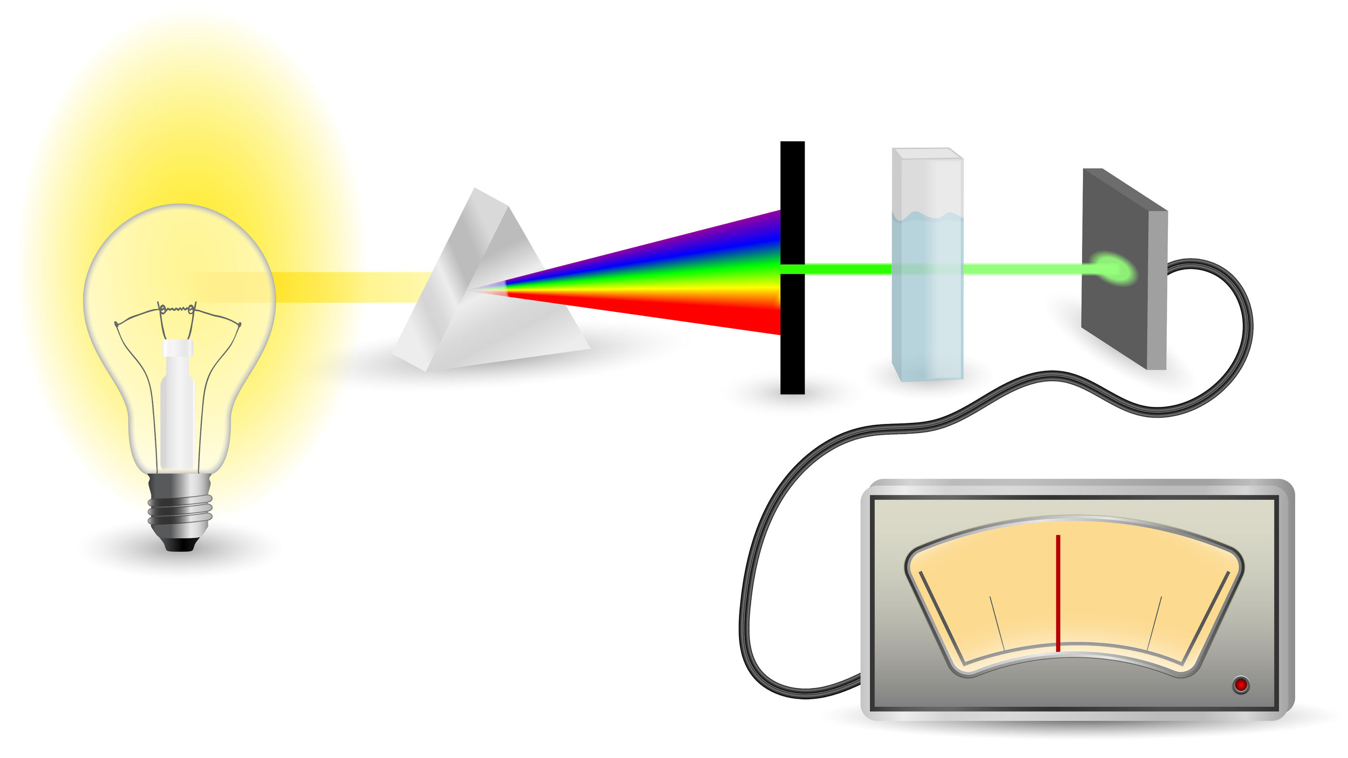
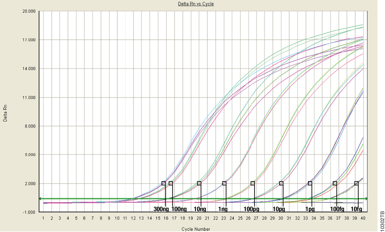
When it comes to nucleic acid quantitation, real-time or quantitative (qPCR) is considered the gold standard because of its unmatched performance in senstivity, specificity and accuracy. qPCR relies on thermal cycling, consisting of repeated cycles of heating an cooling for DNA melting and enzyamtic replication. Detection instrumentation is capable of measuring the accumulation of DNA product after each round of amplification in real time.

Because PCR amplifies specific regions of DNA, the method is highly sensitive, specific to DNA, and it can determine whether a sample is truly able to be amplified. Degraded DNA or free nucleotides, which might otherwise skew your quantiation, will not contribute to the signal, and your measurement will be more accurate.

However, while qPCR does provide technical advantages, the method requires special instrumentation, specialized reagents and is a more time-consuming process. In addition, you will probably need to optimize your qPCR assay for each of your targets to achieve your desired results.

Because of the added complexity and cost, qPCR is a technique suited for post-library quantitation when you need to know the exact amount of amplifiable, adapter-ligated DNA.  PCR is the only method capable of specifically targeting these library constructs over other DNA that may be present. This specificity is important because accurate normalization is especially critical for producing even coverage in multiplex experiments where equimolar amounts of several libraries are added to a pooled sample. This normalization process is essential  if your are screening for rare variants that might be lost in background and go undetected if underrepresented in a mixed pool.


Read Part 1: When Every Step Counts: Quantitation for NGS

Read Part 2: Nucleic Acid Quantitation by UV Absorbance: Not for NGS

Read Part 3: Fluorescence Dye-Based Quantitation: Sensitive and Specific for NGS Applications

ISO 18385: The Creation of a “Forensic Grade” Standard

Today’s blog is from guest blogger Charles Stollberg. Charlie is a Promega production scientist in the Genetic Identity group and is focused on manufacturing inventory material and production process improvements. He’s been with the company for about 4 years. He graduated from UW-Whitewater in 2007 with a bachelor’s degree in cell biology. Prior to Promega, he worked in a small genetics lab studying lethal recessive traits in cattle.

ForensicGradeLogoForensic DNA laboratories rely on reagent and plastics manufacturers to supply high-quality products with minimal interference from contaminating DNA. With the increasing sensitivity of short tandem repeat (STR) amplification systems, levels of DNA that were previously undetected may now generate partial profiles. To address the concern of laboratories worldwide regarding the potential of low-level DNA contamination in consumables, ISO 18385 was developed to provide requirements for minimizing the risk of human DNA contamination events during the manufacturing process. Many of you may not have heard of ISO 18385, so I’d like to give you an introduction to how the standard came to be.

Continue reading “ISO 18385: The Creation of a “Forensic Grade” Standard”

Fluorescence Dye-Based Quantitation: Sensitive and Specific for NGS Applications

This is the third post in a series of blogs on quantitation for NGS applications written by guest blogger Adam Blatter, Product Specialist in Integrated Solutions at Promega.

Fluorescent dye-based quantitation uses specially designed DNA binding compounds that intercalate only with double stranded DNA molecules. When excited by a specific wavelength of light, only dye in the DNA-bound state will fluoresce. These aspects of the technique contribute to low background signal, and therefore the ability to accurately and specifically detect very low quantities of DNA in solution, even the nanogram quantities used in NGS applications.

For commercial NGS systems, such as the Nextera Rapid Capture Enrichment Protocol by Illumina, this specificity and sensitivity of quantitation are critical. The Nextera protocol is optimized for 50ng of total genomic DNA. A higher mass input of genomic DNA can result in incomplete tagmentation, and larger insert sizes, which can adversely affect enrichment. A lower mass input of genomic DNA or low-quality DNA can generate smaller than expected inserts, which can be lost during subsequent cleanup steps, giving lower diversity of inserts.

Continue reading “Fluorescence Dye-Based Quantitation: Sensitive and Specific for NGS Applications”

Nucleic Acid Quantitation by UV Absorbance: Not for NGS

schematic diagram of UV-Vis Absorbance Method
For UV-Vis Spectrophotometry, light is split into its component wavelengths and directed through a solution. Molecules in the solution absorb specific wavelengths of light.

This is the second in a series of four blogs about Quantitation for NGS is written by guest blogger Adam Blatter, Product Specialist in Integrated Solutions at Promega.

Perhaps the most ubiquitous quantitation method is UV-spectrophotometry (also called absorbance spectroscopy). This technique takes advantage of the Beer-Lambert Law: an observation that many compounds absorb UV-Visible light at unique wavelengths, and that for a fixed path length the absorbance of a solution is directly proportional to the concentration of the absorbing species. DNA, for example has a peak absorbance at 260nm (A260nm).

This method is user friendly, quick and easy. But, it has significant limitations, especially when quantitating samples for NGS applications.

Continue reading “Nucleic Acid Quantitation by UV Absorbance: Not for NGS”

When Every Step Counts: Quantitation for NGS

13170MA-800x277This series of blogs about Quantitation for NGS is written by guest blogger Adam Blatter, Product Specialist in Integrated Solutions at Promega.

As sequencing technology races toward ever cheaper, faster and more accurate ways to read entire genomes, we find ourselves able to study biological systems at a level never before possible. From basic science to translational research, massively parallel sequencing (also known as next-generation sequencing or NGS) has opened up new avenues of inquiry in genomics, oncology and ecology.

Many commercial sequencing platforms have been established (e.g., Illumina, IonTorrent, 454, PacBio), and new technologies are developed every day to enable new and unique applications. However, all of these platforms and technologies work off the same general principle: nucleic acid must be extracted from a sample, arranged into platform-specific library constructs, and loaded into the sequencer. Regardless of the sample type or the platform used, every step throughout this workflow is critical for successful results. An often overlooked part of the NGS workflow is sample quantitation. Here we are presenting the first in a series of four short blogs about the critical step of quantitation in NGS workflows.

Sample input is critical to NGS in terms of both quality and quantity. Knowing how much DNA you have, often in nanogram quantities, can make the difference between success and failure. There are several key points in the NGS workflow where sample quantitation is important before you can proceed:

  • After initial nucleic acid extraction from the sample matrix and before proceeding with library preparation
  • Post-library preparation when pooling barcoded libraries for multiplexing
  • Final pooled library quantitation immediately before loading for sequencing

There are several common methods for quantitating nucleic acids: UV-spectroscopy, Fluorescence spectoscopy, real-time quantitative PCR (qPCR). Because of inherent differences in sensitivity, specificity, time and cost, each of these techniques pose certain advantages and disadvantages with respect to the specific sample you are quantitating. Our next three blogs will discuss each of these methods against the backdrop of quantitating samples for NGS applications.


Read Part 2: Nucleic Acid Quantitation by UV Absorbance: Not for NGS

Read Part 3: Fluorescence Dye-Based Quantitation: Sensitive and Specific for NGS Applications

Read Part 4: Real-Time (Quantitative) qPCR for Quantitating Library Prep before NGS

There and Back Again, Part 1

In 2014, Promega created a special incentive to reward field science consultants who help the scientific community take advantage of the our on-site stocking program. The winners had to meet ambitious criteria to receive 2 round-trip tickets to anywhere in the world, a week of paid vacation and spending money. Our four winners from 2014 will share photos and stories about their journeys in a semi-regular Friday feature on the Promega Connections Blog.

Today’s travelogue comes to us from Mica Zaragoza, a senior client rep, who used his award to travel to Australia and New Zealand.

When initially introduced to the ambitious Helix award, I was amazed at the prospect of selecting anywhere in the world to travel, while blogging about my the adventures. Both humbled and amazed to receive this opportunity, my wife and I embarked on a journey across the Pacific.

Hyde Park in Sydney, Australia.

Sydney, Australia

Departing our home in Chicago, my wife Crystal and I started our journey with a 5-hour trip to San Francisco for a layover before the 14 hour journey to Sydney. After jumping into the future (Thurs became Saturday), our first visit was to Central Sydney’s Hyde Park.

Taking jet lag into consideration, we decided to double-down by freshening up and dropping luggage to kick off our day at 7:30am. My first Australian purchase? Coffee! Continue reading “There and Back Again, Part 1”

A Normalization Method for Luciferase Reporter Assays of miRNA-Mediated Regulation

Today’s blog is from guest blogger Ken Doyle of Loquent, LLC. Here, Ken reviews a 2014 paper highlighting specific considerations for using reporter assays to study miRNA-mediated gene regulation.

mirna

The accelerated pace of research into noncoding RNAs has revealed multiple regulatory roles for microRNAs (miRNAs). These diminutive noncoding RNA species—typically 20-24 nucleotides in length—are now known to mediate a broad range of biological functions in plants and animals. In humans, miRNAs have been implicated in various aspects of development, differentiation, and metabolism. They are known to regulate an assortment of genes involved in processes from neuronal development to stem cell division. Dysregulation of miRNA expression is associated with many disease states, including neurodegenerative disorders, cardiovascular disease, and cancer.

Typically, miRNAs act as post-transcriptional repressors of gene expression, either by targeted degradation of messenger RNA (mRNA) or by interfering with mRNA translation. Most miRNAs exert these effects by binding to specific sequences called microRNA response elements (MREs). These sequences are found most often within the 3´-untranslated regions (3´-UTRs) of animal genes, while they may occur within coding sequences in plant genes.

Studies of the regulatory roles played by miRNAs often involve cell-based assays that use a reporter gene system, such as luciferase or green fluorescent protein. In a standard assay, the reporter gene is cloned upstream of the 3´-UTR sequence being studied; this construct is then cotransfected with the miRNA into cells in culture. A study by Campos-Melo et al., published in September 2014, examined this experimental approach for miRNAs from spinal cord tissues, using firefly luciferase as the reporter gene and Renilla luciferase as a transfection control.

Continue reading “A Normalization Method for Luciferase Reporter Assays of miRNA-Mediated Regulation”

Think your budget is too small to start your new lab? Well, think again.


eh62Guest blog by Joanna Stevenson, Promega AG

You are young, dynamic and full of initiative. Your dreams oscillate around setting your playground and working without being told what to do and how to do it. You think it is the only way for you to produce results that others will envy. Well, your dreams can actually come true sooner than you think— with your persistence and with a little help from the Promega New Lab Set Up Program.

Even after receiving for external funding, you probably have a few items you still need to set up your new lab. Maybe you have already established your first lab, but you need to change the location. In any case, maximize you budget and continue dreaming. It doesn’t matter if you are in the USA, Australia, Spain or Switzerland –we can help. Please visit our program at www.promega.com/newlab to find out how.

Avoid False Hits During Compound Screening for Drug Discovery

One goal of drug discovery and research programs is to reduce false hits as early as possible in the process. Follow-up on false hits is costly in terms of time and resources, and the longer the false hits remain in the drug development pipeline, the more costly they are. So methods that can easily reduce the number of false hits during compound screening early in the discovery process are particularly sought after.

Reporter assays have proven to be invaluable tools for elucidating the mechanisms of action of small molecules or other agents on signaling pathways within cells, and the luciferase reporter assay has become a standard research tool in the biological research laboratory.

However, one caveat of using standard luciferase-based reporter assays for larger-scale compound screening efforts is the frequency of false hits that result from direct interaction of compounds with the luciferase reporter. This issue can be mitigated with a “coincidence reporter” system where two independent reporter proteins are produced from a single transcript. In this type of assay, a bicistronic transcript is stoichiometrically translated into two nonhomologous reporters by means of a 2A “ribosomal skipping” sequence. Since it is unlikely that compounds will interact with two distinct types of reporter, “coincident” responses will indicate on-target activity. Such a coincident reporter system provides an important control against costly false hits early in drug discovery research programs.

A paper published online in ACS Chem Biol in February describes the first successful application of the firefly/NanoLuc luciferase coincidence reporter system to identify new pathways that up-regulate PARK2 expression.

Continue reading “Avoid False Hits During Compound Screening for Drug Discovery”