At Promega we like to celebrate the good things, little and big, from the publication of a peer-reviewed paper from a customer or one of our scientists, to the completion of a dissertation defense, to the launch of an exciting new technology. Today we celebrate, along with our local Madison and our global scientific communities, the public opening of our new cGMP manufacturing facility, dedicated to serving customers who need molecular biology reagents for IVD assays.
We could not have asked for a more beautiful fall day for the events. We’ll be live blogging from the opening to share a little bit of the celebration with our Promega Connections community as well.
Promega employees and Wisconsin community members gather for opening ceremonies
The concept of cell death as a normal cell fate was articulated only three years after Schleiden and Schwann introduced the Cell Theory when, in 1874, Vogt described natural cell death as an integral part of toad development (as cited in Cotter and Curtin, 2003). Since these early observations, natural cell death has been described “anew” several times. In 1885 Flemming provided the first morphological description of a natural cell death process, which we now label “apoptosis”, a term coined by Kerr and colleagues to describe the unique morphology associated with a cell death that differs from necrosis (as cited in Kerr et al. 1972).
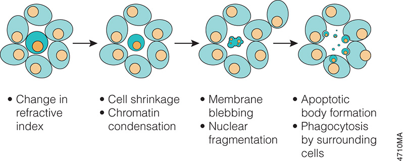
Progression of morphology changes during apoptosis.
In the 1970s and 1980s, studies revealed that apoptosis not only had specific morphological characteristics but that it was also a tightly regulated process with specific biochemical characteristics. Studies of cell lineage in the nematode, Caenorhabditis elegans, showed that apoptosis was a normal feature of the nematode’s invariant developmental program (Hengartner, 1997). At the biochemical level, Wyllie showed that DNA degradation by a specific endonuclease during apoptosis resulted in a DNA ladder composed of mono- and oligonucleosomal-sized fragments (Wyllie, 1980).
These and many other studies have proven that apoptosis is a critical component of development, and when it doesn’t happen appropriately, it can be pathological, leading to cancers or other diseases. Therefore, understanding how and when apoptosis occurs and the many signals that can trigger this process is a focus of many laboratory experiments.
There are many ways to detect apoptosis in cells or tissues. This blog describes some of the most common ones.
Back in 2009, we reported on the problem of cell line contamination (1). In that article we reported the statistics that an estimated 15–20% of the time, the cell lines used by researchers are misidentified or cross-contaminated with another cell line (1). This presents a huge problem for the interpretation of data and the reproducibility of experiments, a key pillar in the process of science. We have revisited this topic several times, highlighting the issues cell and tissue repositories have discovered with cell lines submitted to them (2) and discussing the new guidelines issued by ANSI (3,4) for researchers regarding when during experimental processes cell lines should be authenticated and what methods are acceptable for identifying cell lines.
Just recently two papers were voluntarily retracted by their authors because of cross contamination among cell lines used in the laboratories. The first that came to my attention represented the first retraction from Nature Methods in its nine years of publication. In this paper, cross contamination of a primary gliomasphere cell lines with HEK cells expressing GFP resulted in “unexplained autofluorescence” associated with tumorigenicity (5). The second paper, retracted from Cancer Research by the original authors, was also another cross contamination story involving HEK cells (6). In this story a gene was incorrectly described as a tumor suppressor, that when silenced led to the formation of tumors in nude mice. It turns out that the contaminating HEK cells also failed to express this same gene.
So because of cross contamination of cell lines, two groups have voluntarily retracted papers. Being open and honest about what had happened with the cell lines and reaching the decision to retract the papers could not have been an easy thing, but these decisions benefit the scientific community in many ways. Obviously they benefit the researchers doing work on the specific research questions addressed by the papers by preventing researchers from pursuing paths that lead to dead ends. But in the bigger picture these retractions reinforce the argument that cell line authentication needs to become a routine and accepted part of any experimental process that depends on cell culture if we are to have confidence in the experimental results.
References
Dunham, J.H. and Guthmiller, P. (2009) Doing good science: Authenticating cell line identity. Promega Notes101, 15–18.
I am the mother of a six-year-old girl who loves to get magazines in the mail. For several years my daughter has received an enjoyed popular kids’ science/international culture magazine. The stories are short and simple, and this magazine usually does a good job of presenting factual information in easy-to-digest forms. Each magazine comes with a set of animal cards, which we have diligently collected.
However, the latest issue that came to our mailbox really got me thinking. The final pages featured artwork by the young readers. I love the idea of featuring the work of the readers. Usually, my daughter loves seeing what other children her age from around the world draw and take pictures of, and sometimes we have some pretty interesting discussions about the work.
This time though we didn’t spend much time talking about the art work. She wasn’t particularly interested, and I wasn’t sure I what I thought. But I may have missed a teachable moment. The theme for the pages was a Halloween-minded “spooky science”, and all of the pictures were of “mad scientists” alone at work doing presumably nefarious things in their laboratories. Of the eight drawings pictured, six of them pictured scientists that were human, and five of the humans were male. All of them were pale-skinned. The sole female scientist, whose lab featured a certificate with the words “monster maker”, was drawn by a girl. The ages of the children submitting the work ranged from 9 to 14. Continue reading “Is This What a Scientist Looks Like?”
Hyla versicolor (Copes grey treefrog) Photo credit: LA Dawson wikipedia
Ever since a colleague introduced me to the Spadefoot Toad and the practice of monitoring frog and toad calls during the summer months as one way of tracking the prevalence and health of frogs and toad populations over time, I have been intrigued by any research on frog and toad calling. So, when I saw news headlines about “multitasking” Copes grey treefrogs (Hyla chrysocelis) and their “popularity” among the females of their species, I couldn’t resist.
The paper, published inAnimal Behaviour by Ward and colleagues describes an investigation of the predictions of the “multitasking hypothesis”. Briefly this hypothesis predicts two things. The first prediction states that when you have a signal that has components that negatively covary (in order for one component to occur the second one is reduced), say pitch and volume, there will be a tradeoff between the two things (e.g., I can sing at a really high pitch, but not very loudly). The second prediction is that the individual on the receiving end of this signal is going to prefer the individual who can do both at the same time (e.g., sing at a really high pitch, very loudly). In this study, the authors looked at female frog choice based on three factors: call rate (the number of calls per minute), call duration (number of pulses per call) and call effort (calls/min x pulses/call = pulses/min). They asked two questions: Is there a tradeoff between rate and duration, and do female frogs prefer the male frogs that exhibit the higher overall “call efforts”?
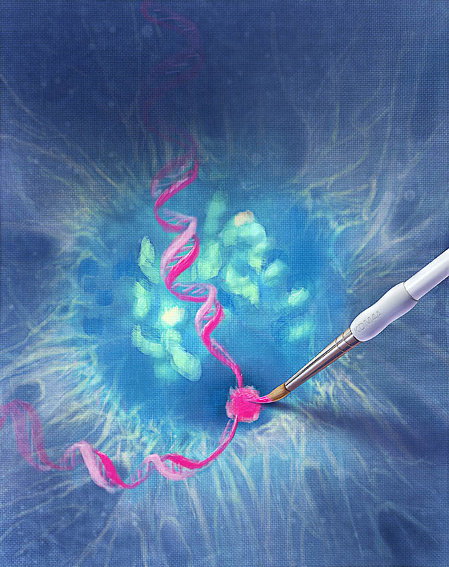
Designed by Nick Klein for ISO-form, courtesy of Promega.
Tumor cells are characterized by many features: including uncontrolled proliferation, to loss of contact inhibition, acquired chromosomal instability and gene copy number changes among them. Some of those copy number changes are site-specific, but very little is known about the mechanisms or proteins involved in creating site-specific copy number changes. In a recently published Cell paper, Black and colleagues, propose a mechanism for site-specific copy number variations involving histone methylation proteins and replication complexes.
Previous work from Klang et al. had shown that local amplification of chromosomal regions occurs during S phase and that chromatin structure plays a critical role in this amplification (2), and other work by Black and colleagues (3) implicated KDM4A in changing timing of replication by altering chromatin accessibility in specific regions. Other research also had shown that KDM4A protein levels influence replication initiation and that KDM4A has a role in some DNA damage response pathways (4,5). Looking at the body of work, Black et al. hypothesized that KDM4A, with its roles in replication, might possibly provide link into the mechanism of site-specific copy number variation in cancer. Continue reading “Site-specific copy number variations in cancer: A story begins to unfold”
Remember the bubble getter? Siliconizing sequencing gel glass plates? Carrying out sequencing reactions in strip tubes? Diagramming, by hand, your cloning scheme and calculating the cut sizes with a hand-held calculator? Marking plates for plaque lifts with india ink?
This video is for all of you who were in the lab when life was “one gene, one graduate student”. What other oldie but goodies can you think of? Leave a comment or tweet @promega #backinmyday
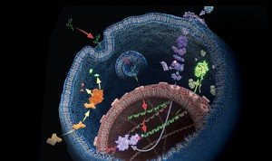
Search the PubMed database for “dual-luciferase” and citations abound. The Dual-Luciferase® Reporter Assay is a powerful tool that allows researchers to ask a multitude of questions about gene control and expression in a system that itself could be normalized and internally controlled. For more than 15 years, firefly and Renilla luciferases have formed the basis of a range of powerful assays and research tools for scientists who are asking questions about the deep and complex genetic and cellular story associated with cancer. Here we talk a bit of about bioluminescent chemistries, some of the newest bioluminescent tools available, and how some of these tools can be used to probe the deeper questions of cell biology, including cancer biology.
The initial paper from the Wild Life in Our Homes study by Dunn et al. found a correlation between the presence of dogs and specific bacterial communities on pillowcases and TV screens.
Back in the fall, I received a sampling kit, an Informed Consent form and instructions for collecting samples for the Wild Life In Our Homes citizen science project. I carefully swabbed the requested surfaces: exterior and interior door trim, kitchen counter tops, pillowcases, etc., and sent my samples in. I later received confirmation that my samples had been received and again later confirmation that they were being analyzed.
The first paper from this project has been published by Dunn et al. in PLOS ONE (Home Life: Factors Structuring the Bacterial Diversity Found within and between Homes). This initial report covers the first 40 homes sampled, all from the Raleigh-Durham, NC, USA area. Volunteers sampled their homes in the Fall of 2011, collecting specimens from nine areas: cutting boards, kitchen counters, refrigerator, toilet seat, pillowcase, door handle, TV screen, and interior and exterior door trim. The scientists used direct PCR and high-throughput sequencing to sequence the bacterial 16S rRNA gene from the submitted samples. By doing this they were able to estimate the diversity within each sample—they did not distinguish between live and dead organisms, and they did not sequence anything other than the bacterial 16SrRNA, so this study is limited to bacteria. Continue reading “About the Wild Life in Our Homes (at least the single-celled kind)”
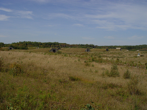
Bunkers at Aroostook National Wildlife Refuge. photo credit: USFWS/Steve Agius
A lot has happened since I first wrote about White-Nose Syndrome, the fungal disease that has devastated bat populations in North America. The disease, caused by the cold-loving fungus Geomyces destructans (now renamed Psuedogymnoascus destructans), has been identified in many more places, including most recently confirmed cases in Georgia, South Carolina, Illinois and Missouri in the United States and Prince Edward Island, Canada.
Controlling the spread of this disease is a tremendous problem, because as I indicated in a previous blog post, keeping a hardy fungus from spreading among a population of densely packed small animals in tiny, cold damp areas is not a simple task.
This problem is going to require creative solutions, and scientists at the U.S. Fish and Wildlife Service may have come up with a great idea that answers two questions: How do you control the spread of White-Nose Syndrome and what do you do with 43 unused Air Force bunkers?
XWe use cookies and similar technologies to make our website work, run analytics, improve our website, and show you personalized content and advertising. Some of these cookies are essential for our website to work. For others, we won’t set them unless you accept them. To learn more about our approach to Privacy we invite you to Read More
By clicking “Accept All”, you consent to the use of ALL the cookies. However you may visit Cookie Settings to provide a controlled consent.
We use cookies and similar technologies to make our website work, run analytics, improve our website, and show you personalized content and advertising. Some of these cookies are essential for our website to work. For others, we won’t set them unless you accept them. To find out more about cookies and how to manage cookies, read our Cookie Policy.
If you are located in the EEA, the United Kingdom, or Switzerland, you can change your settings at any time by clicking Manage Cookie Consent in the footer of our website.
Necessary cookies are absolutely essential for the website to function properly. These cookies ensure basic functionalities and security features of the website, anonymously.
Cookie
Duration
Description
cookielawinfo-checbox-analytics
11 months
This cookie is set by GDPR Cookie Consent plugin. The cookie is used to store the user consent for the cookies in the category "Analytics".
cookielawinfo-checbox-functional
11 months
The cookie is set by GDPR cookie consent to record the user consent for the cookies in the category "Functional".
cookielawinfo-checbox-others
11 months
This cookie is set by GDPR Cookie Consent plugin. The cookie is used to store the user consent for the cookies in the category "Other.
cookielawinfo-checkbox-advertisement
1 year
The cookie is set by GDPR cookie consent to record the user consent for the cookies in the category "Advertisement".
cookielawinfo-checkbox-necessary
11 months
This cookie is set by GDPR Cookie Consent plugin. The cookies is used to store the user consent for the cookies in the category "Necessary".
cookielawinfo-checkbox-performance
11 months
This cookie is set by GDPR Cookie Consent plugin. The cookie is used to store the user consent for the cookies in the category "Performance".
gdpr_status
6 months 2 days
This cookie is set by the provider Media.net. This cookie is used to check the status whether the user has accepted the cookie consent box. It also helps in not showing the cookie consent box upon re-entry to the website.
lang
This cookie is used to store the language preferences of a user to serve up content in that stored language the next time user visit the website.
viewed_cookie_policy
11 months
The cookie is set by the GDPR Cookie Consent plugin and is used to store whether or not user has consented to the use of cookies. It does not store any personal data.
Analytical cookies are used to understand how visitors interact with the website. These cookies help provide information on metrics the number of visitors, bounce rate, traffic source, etc.
Cookie
Duration
Description
SC_ANALYTICS_GLOBAL_COOKIE
10 years
This cookie is associated with Sitecore content and personalization. This cookie is used to identify the repeat visit from a single user. Sitecore will send a persistent session cookie to the web client.
vuid
2 years
This domain of this cookie is owned by Vimeo. This cookie is used by vimeo to collect tracking information. It sets a unique ID to embed videos to the website.
WMF-Last-Access
1 month 18 hours 24 minutes
This cookie is used to calculate unique devices accessing the website.
_ga
2 years
This cookie is installed by Google Analytics. The cookie is used to calculate visitor, session, campaign data and keep track of site usage for the site's analytics report. The cookies store information anonymously and assign a randomly generated number to identify unique visitors.
_gid
1 day
This cookie is installed by Google Analytics. The cookie is used to store information of how visitors use a website and helps in creating an analytics report of how the website is doing. The data collected including the number visitors, the source where they have come from, and the pages visted in an anonymous form.
Advertisement cookies are used to provide visitors with relevant ads and marketing campaigns. These cookies track visitors across websites and collect information to provide customized ads.
Cookie
Duration
Description
IDE
1 year 24 days
Used by Google DoubleClick and stores information about how the user uses the website and any other advertisement before visiting the website. This is used to present users with ads that are relevant to them according to the user profile.
test_cookie
15 minutes
This cookie is set by doubleclick.net. The purpose of the cookie is to determine if the user's browser supports cookies.
VISITOR_INFO1_LIVE
5 months 27 days
This cookie is set by Youtube. Used to track the information of the embedded YouTube videos on a website.
Performance cookies are used to understand and analyze the key performance indexes of the website which helps in delivering a better user experience for the visitors.
Cookie
Duration
Description
YSC
session
This cookies is set by Youtube and is used to track the views of embedded videos.
_gat_UA-62336821-1
1 minute
This is a pattern type cookie set by Google Analytics, where the pattern element on the name contains the unique identity number of the account or website it relates to. It appears to be a variation of the _gat cookie which is used to limit the amount of data recorded by Google on high traffic volume websites.


 Remember the bubble getter? Siliconizing sequencing gel glass plates? Carrying out sequencing reactions in strip tubes? Diagramming, by hand, your cloning scheme and calculating the cut sizes with a hand-held calculator? Marking plates for plaque lifts with india ink?