Monitoring Mass Spec Instrument Performance and Sample Preparation

Proteomics, the analysis of the entire protein content of a living system, has become a vital part of life science research, and mass spectrometry (MS) is the method for analyzing proteins.  MS analysis of protein content allows researchers to identify proteins, sequence them and determine the nature of post translational modifications.

LC/MS performance monitoring. Each run used 1μg of human predigested protein extract injected into the instrument (Waters NanoAquity HPLC System interfaced to a Thermo Fisher Q Exactive™ Hybrid Quadrupole-Orbitrap Mass Spectrometer). Peptides were resolved with a 2-hour gradient. Weekly monitoring with the human extract ensured consistent analytical performance of the instrument.

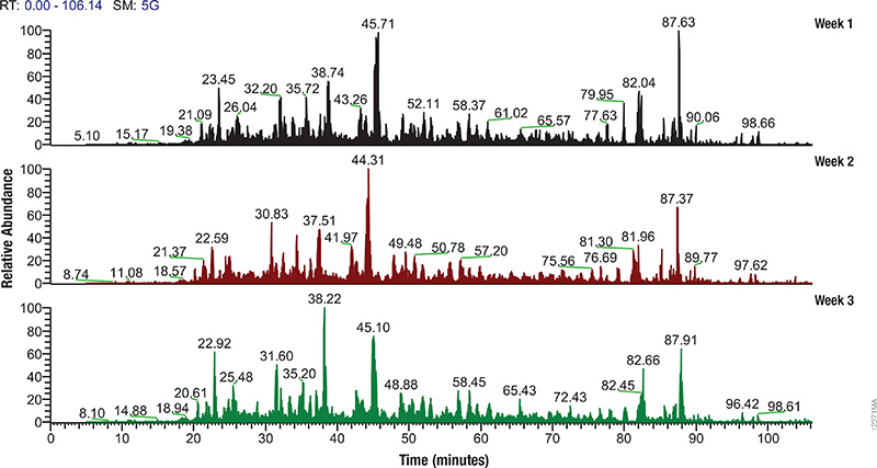
Mass spectrometry allows characterization of molecules by converting them to ions so that they can be manipulated in electrical and magnetic fields. Basically a small sample (analyte) is ionized, usually to cations by loss of an electron. After ionization, the charged particles (ions) are separated by mass and charge;  the separated particles are measured and data displayed as a mass spectrum. The mass spectrum is typically presented as a bar graph where each peak represents a single charged particle having a specific mass-to-charge (m/z) ratio. The height of the peak represents the relative abundance of the particle. The number and relative abundance of the ions reveal how different parts of the molecule relate to each other.

For the study of large, organic macromolecules, matrix associated laser desorption/ionization (MALDI) or tandem mass spec/collision induced dissociation (MS/MS) techniques are often used to generate the charged particles from the analyte. MS analysis brings sensitivity and specificity to proteome analysis. The technique has excellent resolution and is able to distinguish one ion from another, even when their m/z ratios are similar. Macromolecules are present in extremely different concentrations in the cells, and MS analysis can detect biomolecules across five logs of concentration.

Continue reading “Monitoring Mass Spec Instrument Performance and Sample Preparation”

Shedding Light on Protein:Protein Interactions with NanoBRET™ Technique

NanoBRET™ TechnologyIf you are trying to investigate protein:protein interactions inside cells, you know how important physiologically relevant results are. If you overload your cells with fusion constructs, your protein interactions may not actually reflect what is going on in the cell, and if your BRET energy donor and acceptor do not have sufficiently separated spectra, you can pick up a fair amount of noise in your experiment. Using the new superbright NanoLuc® Luciferase, and the HaloTag® Technology, we have developed a sensitive BRET system to help you take a better look specific protein interactions that interest you. Promega research scientist, Danette Daniels, describes the system in the Chalk Talk below:

Plant Biologists Take the Lead on Elucidating Zombie Genetics

claimtoken-5367ae87b7cd2

Biology is full of stories that read like a modern day zombie apocalypse. For instance, the parasite Toxoplasma gondii has been in the news for its ability to infect the brains of rats, and reprogram their normal behavioral responses such that they lose their innate fear of cats. Previously, we reviewed the research about the parasitic fungus that infects ants, causing drastic changes in typical ant behavior to aid in distribution of the fungal spores.

In April of this year, MacLean and colleagues published research in PLOS Biology describing interactions between a phytoplasma parasite and Arabidopsis thaliana. What is nice about this particular “zombie” biology story is that the researchers present the beginnings of the genetics that underlie the plant-parasite-insect relationship, moving beyond a description of the phenotypic changes that occur to describing an actual mechanism for those changes.

Continue reading “Plant Biologists Take the Lead on Elucidating Zombie Genetics”

Ghost of the Himalayas (Snow Leopard) Caught on Camera In a Successful Hunt

Snow LeopardPaul Steyn has posted an amazing series of photographs taken by Adam Riley in Hemis National Park in the Himalayas on the National Geographic News web site. These photographs are the  first photo documentation of a successful snow leopard hunt, and underscore the amazing biology of this area. If you are interested in learning more, we have another blog post about early work to isolate induced pluripotent stem cells from adult animals–an attempt to compliment the habitat preservation and other efforts to save this incredible hunter before it truly becomes a ghost.

Butterfly Heal Thyself: What We Can Learn from Self-Medicating Animals in Nature

Can animals actively seek out a plant to heal a parasitic infection or to numb pain? For years scientists have presumed that such activity was restricted to animals capable of more complex cognitive abilities, like chimpanzees. However scientists have described incidents of fruit flies (1), caterpillars (2)  and butterflies  (3) “self medicating” to prevent or cure parasitic infections, suggesting that such behavior may be both innate as well as learned.

Continue reading “Butterfly Heal Thyself: What We Can Learn from Self-Medicating Animals in Nature”

Using NanoLuc® Luciferase to Study Interactions between Environmental Flavobacteria and Mosquitos

nanoluc

Studies of the larval stages of Aedes triseriatus  (Eastern TreeHole Mosquito) indicate that the “tree hole” habitats in which these larva develop contain diverse microflora including the flavobacteria Elizabethkingia and Chryseobacterium. Extracts from these bacteria have many properties that might affect mosquito health, including antibacterial and anti-fungal activities. Understanding how these bacteria affect larval mosquito development has the potential to inform strategies for mosquito control.

Some initial work has been done by expressing Bacillus larvacidal toxins in some species of Gram-negative bacteria. However, only limited success was achieved using laboratory bacterial strains for such studies. Using environmental flavobacteria might prove to be a more useful approach. However, few molecular tools exist to study environmental flavobacteria. GFP reporters have been used to look at larval feeding, but autofluorescence in the pupae limit the usefulness of GFP-labeled strains for quantitative studies. Furthermore, environmental flavobacteria have unique transcription and translation machinery, and selectable markers and expression plasmids from proteobacteria do not function in these wild strains.

Chen and colleagues set out to generate molecular tools to study Flavobacterium hibernum, a fast-growing bacterium from native mosquito habitats. Their goal was to use these tools to see if A. triseratus larvae ingest and digest these bacteria and to test whether or not F. hibernum can be used to as a vector for larvacidal toxins directed against mosquito larvae. The results of their work were published in Applied and Environmental  Microbiology .

To develop a reporter that avoided issues of autofluorescence background for quantitative studies on the feeding behavior, the researchers turned to NanoLuc® Luciferase, a small, bright luciferase derived from the sea shrimp Oplopphorus gracilirostris. This luciferase has been used in mammalian cells for many kinds of studies, but it has not been used as a reporter in bacterial cells prior to the work of Chen et al. They also looked at work with laboratory flavobacteria strains that used a promoter of outer membrane protein A (PompA) to drive reporter expression as a potential system that might also work with environmental flavobacteria strains.

Continue reading “Using NanoLuc® Luciferase to Study Interactions between Environmental Flavobacteria and Mosquitos”

Sustainable Growth: The Edible Kind

community-garden-thumb

From time to time, we use the Promega Connections blog to tell you a little bit more about life here at Promega Corporation. For more than 40 years Promega has consistently integrated the values of corporate responsibility and sustainable business practices in all aspects of our corporate culture and activity; one of those aspects can be found in the Promega cafeterias. As an employee of Promega, one of the things I have considered a key perk is the wonderful menu offerings we have here at the Madison campus. The kitchens offer a varied and fun menu full of healthy choices, using local, seasonal foods as much as possible. The Promega Culinary Garden was covers more than an acre and allows us to grow many of our own vegetables and herbs as well as compost food waste.

Below is a short video highlighting the Culinary Garden Program

For information on other corporate responsibility and sustainable business practices at Promega Corporation, explore our Corporate Responsibility Web site.

Top 10 Innovator—Two Years Running

The ADCC Reporter Bioassays were named a Top 10 innovation by The Scientist Magazine.
The ADCC Reporter Bioassay systems were named a Top 10 innovation by The Scientist Magazine.
For the second year running a Promega technology has made The Scientist Magazine’s list of Top 10 Innovations. Last year it was the NanoLuc® luciferase technology; this year it is the ADCC Reporter Bioassay.

Antibody-dependent cell-mediated cytotoxicity (ADCC) is the main mechanism of action (MOA) of antibodies through which virus-infected or other diseased cells are targeted for destruction by components of the cell-mediated immune system. ADCC assays are often used to assess the effectiveness of monoclonal antibody therapies during the manufacture and development of biologic drugs. The bioluminescent ADCC Reporter Bioassays use an alternative readout at an earlier point in ADCC MOA pathway for the quantification of Fc effector function of antibody-based molecules: the activation of gene transcription through the NFAT (nuclear factor of activated T-cells) pathway in the effector cell.

The bioassay uses engineered Jurkat cells stably expressing the FcγRIIIa receptor, V158 (high affinity) variant, and an NFAT response element driving expression of firefly luciferase. The assay is ADCC MOA-based and features frozen, thaw-and-use effector cells and optimized reagents and protocol to perform a reporter-based ADCC bioassay in a single day. The ADCC Reporter Bioassay correlates with classic cytotoxic ADCC assays and is a suitable replacement for these cumbersome and highly variable assays.

The novel bioassay is linear, accurate, precise and stability indicating. Moreover, the bioassay shows good linear correlation between levels of glycosylation or fucosylation and ADCC activity. All of these features indicate the assay is suitable for use across biologic drug development programs.

Resources for the ADCC Reporter Bioassays:

Piecing the Puzzle Together: Using Multiple Assays to Better Understand What Is Happening with Your Cells

You often need several pieces of information to really understand what is happening within a cell or population of cells. If your cells are not proliferating, are they dying? Or, are you seeing cytostasis? If they are dying, what is the mechanism? Is it apoptosis or necrosis? If you are seeing apoptosis, what is the pathway: intrinsic or extrinsic?

If you are measuring expression of a reporter gene and you see a decrease in expression, is that decrease due to transfection inefficiencies, cytotoxicity, or true down regulation of your reporter gene?

To investigate these multiple parameters, you can run assays in parallel, but that requires more sample, and sample isn’t always abundant.

Multiplexing assays allows you to obtain information about multiple parameters or events (e.g., reporter gene expression and cell viability; caspase-3 activity and cell viability) from a single sample. Multiplexing saves sample, saves time and gives you a more complete picture of the biology that is happening with your experimental sample.

What information do you need about your cells to complete the picture?

Multiplexing assay reagents to measure biomarkers in the same sample has often been considered an application only accomplished with antibodies or dyes and sophisticated detection instrumentation. However, Promega has developed microwell plate based assays for cells in culture that allow multiplexed detection of biomarkers in the same sample well using standard multimode multiwell plate readers. Continue reading “Piecing the Puzzle Together: Using Multiple Assays to Better Understand What Is Happening with Your Cells”

Technology and Life in the Lab

It’s a brave new world in the laboratory, mobile devices, smart phones and tablet computers have transformed the way we do science. We’ve come a long way from “bubble getters” for acrylamide gels and foam bath floats. Today’s laboratory tools come with bells and whistles, touch screens and timers.
ehsept2013Need to design a double restriction enzyme digest? Just whip out your iOS or Android device and check the restriction enzyme tool on the Promega App–no more piles of biotech catalogs on the lab bench. Need to calculate the Tm of an oligo? Or find the recipe for a common buffer? Put away your scientific calculators and your big red protocol books; all you need is a smart phone. Need to count colonies? Forget the counting pen, use your camera phone instead. It’s amazing the tools that we have at our fingertips in the lab. Which ones are your favorites? What tool do you wish you had at your for your lab work? What do you still do with paper and pencil, because frankly nothing works better?