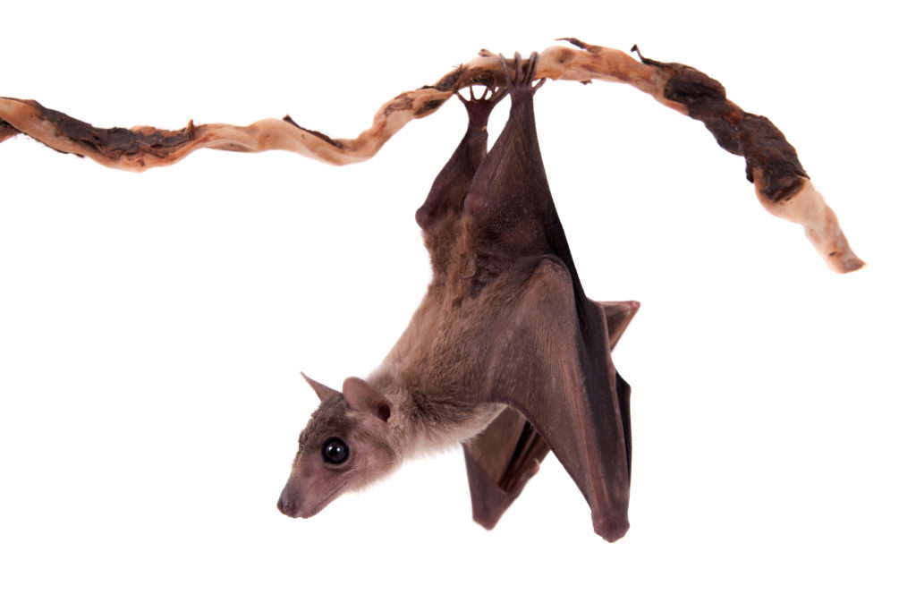
Ebola virus (EBOV) and Marburg virus (MARV) are two closely-related viruses in the family Filoviridae. Filoviruses are often pathogenic, causing hemorrhagic fever disease in human hosts. The Ebola outbreak of 2014 caught the world by surprise by spreading so quickly and severely that public health organizations were unprepared. The devastating outcome was a total of over 11,000 deaths by the time the outbreak ended in 2016. Research that provides further understanding of filoviruses and their potential for transmission is important in preventing future outbreaks from occurring. But what if the outbreak comes from a virus we’ve never seen before?

All in the viral family
A recent study published in the journal Nature Microbiology provides evidence of a newly identified filovirus species. Using serum samples taken from bats, a well-known host for filoviruses, Yang et al. isolated and identified viral RNA for an unclassified viral genome sequence using next generation sequencing analysis. This new virus genome sequence was organized with the same open reading frames as other filoviruses, encoding for nucleoprotein (NP), viral protein 35 (VP35), VP40, glycoprotein (GP), VP30, VP24, and RNA-dependent RNA polymerase (L). This new genome sequence shared up to 54% of the nucleotide sequences for the filovirus species Lloviu virus (LLOV), EBOV and MARV, with MARV being the most similar. Their analysis suggested that this novel virus should be classified within the Filoviridae family tree as a separate genus, Dianlovirus, and was named Měnglà virus (MLAV).
Continue reading “Meet Měnglà Virus: the newest cousin in the Ebola and Marburg virus family tree”
 When someone is admitted to a hospital for an illness, the hope is that medical care and treatment will help them them feel better. However, nosocomial infections—infections acquired in a health-care setting—are becoming more prevalent and are associated with an increased mortality rate worldwide. This is largely due to the misuse of antibiotics, allowing some bacteria to become resistant. Furthermore, when an antibiotic wipes out the “good” bacteria that comprise the human microbiome, it leaves a patient vulnerable to opportunistic infections that take advantage of disruptions to the gut microbiota.