Moving Out of the Cell: Advantages of Cell-Free Protein Expression

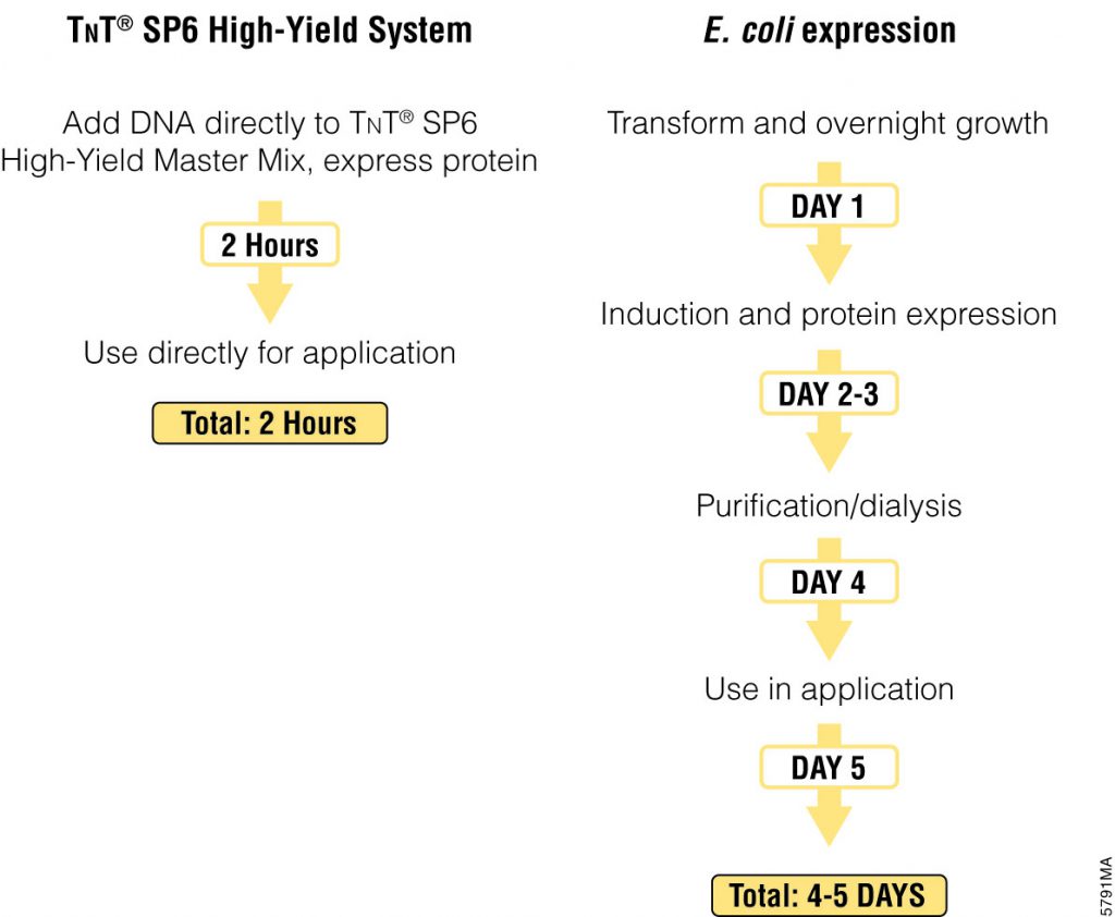
Cell-free protein expression is a simplified and accelerated avenue for the transcription and/or translation of a specific protein in a quasi cell environment. An alternative to slower, more cumbersome cell-based methods, cell-free protein expression methods are simple and fast and can overcome toxicity and solubility issues sometimes experienced in the traditional E. coli expression systems.

Cell-free protein expression offers significant time savings over cell-based expression methods.

Cell-free protein expression offers a convenient method for generating small amounts of protein for a variety of applications (e.g., protein:protein interactions, protein: nucleic acid interactions, structural analysis, functional assays and toxicity screening). This approach lends itself to specific protein labeling with fluorescence, biotin, radioactivity or heavy atoms, via modified charged tRNA’s or amino acids. Cell-free protein expression systems provide quick access to proteins of interest and remain a staple in the collection of tools available for the elucidation of protein structure and function, understanding cellular pathways and mechanisms and high-throughput screening of compounds for drug discovery. There are a number of different cell-free expressions systems, each with different strengths. Deciding which one is right for you depends upon your research needs and goals.

Continue reading “Moving Out of the Cell: Advantages of Cell-Free Protein Expression”

Easy Automated Genomic DNA Isolation for GMO Testing: From Vision to Reality

29980616-July25-PureFood---Kelly-600x300

The European Union (EU) has a zero tolerance policy for products containing any material from non-authorized genetically modified (GM) crops. Seed entering EU markets may not contain even trace amounts of non-authorized genetically modified material. In 2012, as the global use of GM crops increased, seed testing loads in the EU continued to build. Isolating genomic DNA (gDNA) using traditional manual methods was becoming impractical in the face of increasing amounts of material that required testing. There was a growing need for an automated method to isolate gDNA from seed samples. Working to address this need, a group of scientists from the Bavarian Health and Food Safety Authority collaborated with scientists from Promega Corporation to evaluate the Maxwell® 16 Instrument and the associated chemistry as possible a solution for the testing labs.

Continue reading “Easy Automated Genomic DNA Isolation for GMO Testing: From Vision to Reality”

Looking Back: Seeing the Science of My Childhood

Gene silencingScience is all around us— in everything we touch, smell, taste and see. It is in the flowers in our gardens, the molecules of pollen and oils that give those flowers scent, the crystals of sodium chloride that gives our food flavor and the way light is bent and changed to give our world color. There is science in the way we look like our great-great grandmother, and science in the way we are so different from each other. As the granddaughter of a forester and a botanist and the daughter of a science teacher, there has been science in my life for as long as I can remember. Recently my parents moved to a retirement home, and as I spent time helping them downsize, I took pictures of some of the ‘science’ that surrounded my as I grew up.
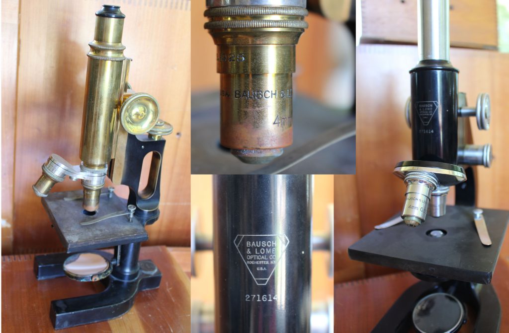
To start, there is “old brassy”, the first microscope I ever used. This microscope, and it’s slightly more modern cousin held places of honor on shelves in my father’s den.microscopes

Held in wooden boxes next to the microscopes were test tubes containing all sorts of mysterious things, including samples gathered by my grandfather while he was a forester in Louisiana. Continue reading “Looking Back: Seeing the Science of My Childhood”

In the Moment with Promega Software Designer, Dave Romanin

26062334-portrait-WEBWhen Dave Romanin came to work for Promega he was fresh out of school with a degree in bacteriology. His plan was to work for a year in manufacturing and then go back to graduate school. But in the end, he didn’t go. There was no incentive, he explains, for him to spend five years in graduate school making little to no money. He didn’t want to write grants or run his own lab, and he enjoyed what he was doing.
Twenty‐four years later, Dave is still here. He’s moved around a bit, first manufacturing, then dispensing, kit packaging and then on to software development with Lou Mezei. Their first software project was a quality control software to capture data from the scales weighing bottles to ensure they were filled correctly. His experience in manufacturing helped him understand what the program needed to do and helped him define the specifications for the software for the programmer. He has been designing software for the last 10 years, and has worked on projects for everyone from marketing to manufacturing.

He describes his job, in part, as a game of cat and mouse. Dave spends hours testing the software, trying to find the weaknesses the developer didn’t anticipate—in essence, trying to break it. When he finds something that throws the software off or causes it to crash, he and the programmer decide on the next steps. Sometimes it is an easy fix, and sometimes they have to decide if it is worth what it would take to fix it. Would a user be likely to ever do what Dave did? Continue reading “In the Moment with Promega Software Designer, Dave Romanin”

Congenital Cytomegalovirus: The Most Common Congenital Infection That You Have Never Heard Of

infant

Down Syndrome. Fetal alcohol syndrome. Spina bifida. Most people have heard of these well know congenital conditions, and know at least in a general sense that they have profound and lasting effects on the children born with them as well as their families. Unfortunately, people are much less aware of a congenital infection that is more common that any of these and affects more infants than all three of the conditions listed above. In fact, this congenital infection causes more cases of congenital disease than all of the 29 conditions currently screened for in most American states combined (1; 2), and yet it is not widely known by the general public. Cytomegalovirus, or CMV, is the most common congenital viral infection in developed countries (3; 4), and the leading cause of congenital sensorineural hearing loss and psychomotor retardation (1).

Continue reading “Congenital Cytomegalovirus: The Most Common Congenital Infection That You Have Never Heard Of”

Inflammasomes: Peeking Inside the Inflammatory Process

Most of us have experienced an inflammatory response at some point in our lives. Fever, achy joints, swelling around a scrape or cut, all of these are forms of inflammatory response. Inflammation is the body’s response to infection or tissue damage and acts to limit harm to the rest of the body. A key player in the inflammation process is a group of protein complexes call inflammasomes. The term “inflammasome” was first used in 2002 by researchers in Switzerland (1) to refer to a caspase-activating protein complex. We now know that inflammasomes are cytosolic multiprotein platforms that assemble in response to pathogens and other signals. Inflammasome assembly results in the processing of the inactive procaspase-1 into the active cysteine-protease enzyme, caspase-1. Caspase-1, in turn, activates the proinflammatory cytokines Interleukins IL-1β and IL-18. In addition, caspase-1 is also required for pyroptosis, which is an inflammatory form of cell death that combines the characteristics of apoptosis (fragmented DNA) and necrosis (inflammation and cytokine release) and is frequently associated with microbial infections.

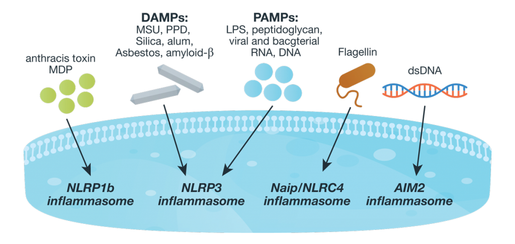
Inflammasome complexes are made up of scaffolding sensor proteins (NLR, AIM2, ALR), and an adaptor protein containing a caspase activation and retention domain (CARD) and inactive procaspase-1. Most inflammasomes are formed with one or two NLRs (NOD-like receptors). However, non-NLR proteins such as AIM2 (absent in melanoma 2) and pyrin can also form inflammasomes. The different sensor proteins are activated by different types of outside stimuli, and inflammasomes are loosely sorted into families based on the protein forming these sensors.26061534-Inflamasome-Assay-Blog-figure-WEB Continue reading “Inflammasomes: Peeking Inside the Inflammatory Process”

Top 10 Tips to Improve Your qPCR or RT-qPCR Assays

headache

Scientists have had a love-hate relationship with PCR amplification for decades. Real-time or quantitative PCR (qPCR) can be an amazingly powerful tool, but just like traditional PCR, it can be quite frustrating. There are several parameters that can influence the success of your PCR assay. We’ve highlighted ten things to consider when trying to improve your qPCR results.

Continue reading “Top 10 Tips to Improve Your qPCR or RT-qPCR Assays”

Circulating Biomarkers and Their Applications in Gastric Cancer

cancer cellCancer. It has been the nemesis of medical science for decades. We declare war on it, we wax philosophical about finding a cure for it. We talk about it as if it were a single enemy, but it isn’t—Cancer is not a disease, it is hundreds of diseases. These diseases manifest in every region of the human body. Many cancers, if diagnosed early, can be treated successfully. Unfortunately, there are many forms of cancers that have no external signs and very few symptoms at the early stages. Such is the enemy we face: we know what it is, we know we need to kill it early and in many cases we know how to do it, what we don’t know is how to catch it early.

Gastric cancer kills approximately 745,000 people a year worldwide, making it the third most common cause of cancer-related deaths (1). It has such a high mortality rate because usually it is not detected until the disease has progressed to the later stages (IIIA–IV; 2). When detected this late, the 5-year survival rate ranges from 7–27%, with the median survival being less than 12 months (2). In contrast, when diagnosed early (i.e., cancer that is limited to the submucosal layer) it is curable with an endoscopic mucosal dissection or a minimally invasive surgery. The difference between these two outcomes is time. The earlier the cancer is detected, the better the prognosis.

Currently, upper endoscopy is the primary screening technique for detecting precancerous lesions as well as gastric cancer in the early stages. This technique has a number of downsides: it is invasive, it can have serious side effects (although these are uncommon), and it is expensive and highly dependent on the skill of the endoscopist.  For these reasons, endoscopy screening is likely to suffer from poor participation rates. In addition, endoscopy is not a practical approach in low-income countries. There is clearly a need for a less invasive, sensitive screening test that will detect gastric cancer at an early stage. Continue reading “Circulating Biomarkers and Their Applications in Gastric Cancer”

Interrogating Protein Interactions: An Infographic for NanoBRET™ Assay Design

Yesterday my fellow blogger, Kari, posted a review of the ACS Chemical Biology paper describing a new BRET platform for analyzing protein-protein interactions. If you are interested in studying induction and inhibition of protein interactions in real time, take a look at the infographic below to learn how to develop a NanoBRET™ Assay to monitor your protein of interest.

21421668-IG-CR-NanoBRET-Infographic-Final

Characterizing Immune-Modulating Antibodies Using Bioluminescence

Immune checkpoint pathways such as PD-1/PD-L1 and CTLA-4 are promising new immunotherapy targets for the treatment of cancer and autoimmunity. Immune checkpoint reporter-based bioassays provide a simple, consistent, and reliable cell-based assay to measure Ab function throughout the drug development pipeline.

The brief chalk talk below describes the assay principals of the reporter-based bioassay that monitors the functional blockade of PD-1/PD-L1 interactions.