Your Brain on COVID-19: Neurotropic Properties of the SARS-CoV-2 Virus

Updated 4/29/2022 by AnnaKay Kruger

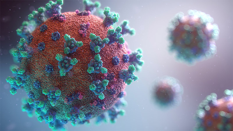
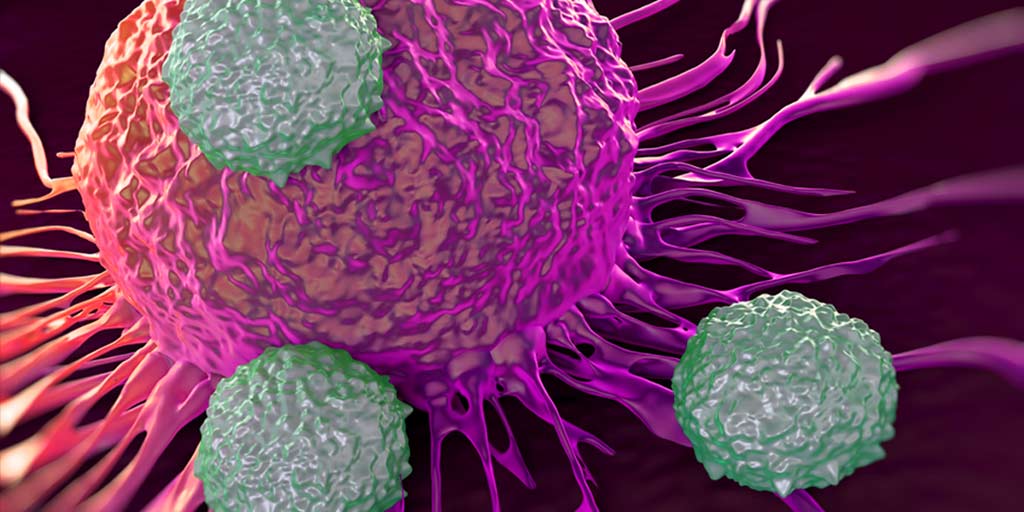
Artist conception of coronavirus in the brain. Researchers are investigating the neurotropic effects of SARS-CoV-2

Viruses are both fascinating and terrifying. Stealthy, insidious and often deadly, they turn our own cells against us. Over the past year, we have all had a firsthand view of what a new and unknown virus can do. The SARS-CoV-2 virus has caused a global pandemic, and left scientists and medical professionals scrambling to unravel its mysteries and find ways to stop it.

COVID-19 is considered a respiratory disease, but we know that the SARS-CoV-2 virus can affect other systems in the body including the vascular and central nervous systems. In fact, some of the most noted symptoms of SARS-CoV-2 infection, headache, and the loss of the sense of taste and smell, are neurological— not respiratory— symptoms.

Continue reading “Your Brain on COVID-19: Neurotropic Properties of the SARS-CoV-2 Virus”

Increasing Testing Efficiency with Multiplexed Detection of SARS-CoV-2 and Influenza A and B

In the Northern hemisphere, the cold and flu season is about to start. Most years that means people schedule flu shots, dust off chicken soup recipes and stock up on tissues. If they start to feel sick, they stay home for a day or two, drink hot tea, eat warm soup and—for the most part— go on with their lives. 

This is not, however, most years. This year the world is battling a pandemic virus, SARS-CoV-2. Symptoms of COVID-19, the disease caused by this virus, mirror those of the flu and common cold, and that overlap in symptoms is going to make life more complicated. Most years, a mild cough or minor body aches wouldn’t even warrant a call to the doctor. This year these, and other undiagnosed cold- and flu-like symptoms, won’t be easily ignored. They could mean kids have to stay home from school, and adults have to self-quarantine from work, for up to 2 weeks. In years past people might have been comfortable treating their symptoms at home, this year people will want answers: Is it the flu? Or is it COVID-19?

Continue reading “Increasing Testing Efficiency with Multiplexed Detection of SARS-CoV-2 and Influenza A and B”

Lessons in History, Hope and Living with Lynch Syndrome from the “Daughter of Family G”

Lynch Syndrome is the autosomal dominant hereditary predisposition to develop colorectal cancer and certain other cancers. This simple, one sentence definition seems woefully inadequate considering the human toll this condition has inflicted on the families that have it in their genetic pedigree.

They Called it a Curse

To one family, perhaps the family when it comes to this condition, Lynch Syndrome has meant heartache and hope; grief and joy; death and life. Their story is told by Ami McKay in her book Daughter of Family G, and it is at once both a memoir of a Lynch Syndrome previvor (someone with a Lynch Syndrome genomic mutation who has not yet developed cancer) and a poignant and honest account of the family that helped science put name to a curse.

“The doctors called it cancer. I say it’s a curse. I wish I knew what we did to deserve it.”

Anna Haab from Daughter of Family G (1)

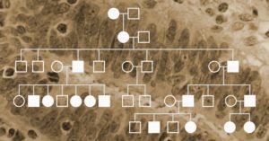
The scientific community first met “Family G” as the meticulously created family tree, filled with the stunted branches that mark early deaths by cancer. The pedigree was first published in 1913 in Archives of Internal Medicine (2). In the article, Dr. Alderd Warthin wrote: “A marked susceptibility to carcinoma exists in the case of certain family generations and family groups.” In 1925, an expanded pedigree of circles and squares was published in Dr. Warthin’s follow up study in the Journal of Cancer Research (3).  But each circle and square in that pedigree denotes a person. Each line represents their dreams together for the future, and Ms. McKay wants us to know their names: Johannes and Anna, Kathrina, Elmer, Tillie, Sarah Anne (Sally); and—most importantly—Pauline. Because without Pauline there would be no story.

Continue reading “Lessons in History, Hope and Living with Lynch Syndrome from the “Daughter of Family G””

Promega Leverages Long-Time Experience in MSI Detection with European Launch of CE-Marked IVD Assay for Microsatellite Instability

The genetic abnormality called microsatellite instability, or MSI, has been linked to cancer since its discovery in 1993 (1). MSI is the accumulation of insertion or deletion errors at microsatellite repeat sequences in cancer cells and results from a functional deficiency within one or more major DNA mismatch repair proteins (dMMR).  This deficiency, and the resulting genetic instability, is closely related to the carcinogenicity of tumors (2).

Historically MSI has been used to screen for Lynch Syndrome, a dominant hereditary cancer propensity. More recently, tumors with deficient MMR function have been identified as being more likely to respond to immune checkpoint inhibitor (ICI) therapies (3.). Because MSI can be the first evidence of an MMR deficiency, MSI-High status is predictive of a positive response to immunotherapies such as ICI therapies. (3).

Learn more about MSI in this short animation.
Continue reading “Promega Leverages Long-Time Experience in MSI Detection with European Launch of CE-Marked IVD Assay for Microsatellite Instability”

RT-qPCR and qPCR Assays—Detecting Viruses and Beyond

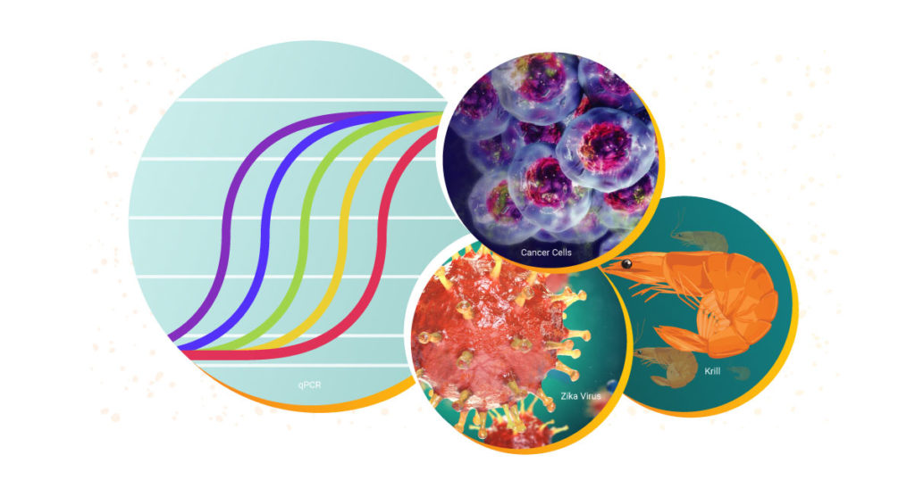
We have all been hearing a lot about RT-PCR, rRT-PCR and RT-qPCR lately, and for good reason. Real-Time Reverse Transcriptase Polymerase Chain Reaction (rRT-PCR) is the technique used in by the Center for Disease Control (CDC) to test for COVID-19. Real-time RT-PCR, or quantitative RT-PCR (RT-qPCR)*, is a specialized PCR technique that visualizes the amplification of the target sequence as it happens (in real-time) and allows you to measure the amount of starting target material in your reaction. You can read more about the basics of this technique, and watch a webinar here. For more about RT-PCR for COVID-19 testing, read this blog.

Both qPCR and RT-qPCR are powerful tools for scientists to have at their disposal. These fundamental techniques are used to study biological processes in a wide range of areas. Over the decades, Promega has supported researchers with RT-qPCR and qPCR reagents and systems to study everything from diseases like COVID-19 and cancer to viruses in elephants and the circadian rhythm of krill.  

Continue reading “RT-qPCR and qPCR Assays—Detecting Viruses and Beyond”

Optimizing PCR: One Scientist’s Not So Fond Memories

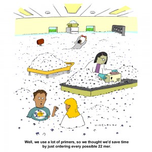
primer_tubesThe first time I performed PCR was in 1992. I was finishing my Bachelors in Genetics and had an independent study project in a population genetics laboratory. My task was to try using a new technique, RAPD PCR, to distinguish clonal populations of the sea anemone, Metridium senile. These creatures can reproduce both sexually and asexually, which can make population genetics studies challenging. My professor was looking for a relatively simple method to identify individuals who were genetically identical (i.e., potential clones).

PCR was still in its infancy. No one in my lab had ever tried it before, and the department had one thermal cycler, which was located in a building across the street. We had a paper describing RAPD PCR for population work, so we ordered primers and Taq DNA polymerase and set about grinding up bits of frozen sea anemone to isolate the DNA. [The grinding process had to be done using a mortar and pestle seated in a bath of liquid nitrogen because the tissue had to remain frozen. If it thawed it became a disgusting mass of goo that was useless—but that is a topic for a different blog.] Since I had never done any of the procedures before, my professor and I assembled the first set of reactions together. When we ran our results on a gel, we had all sorts of bands—just what he was hoping to see. Unfortunately, we realized that we had added 10X more Taq DNA polymerase than we should have used. I repeated the amplification with the correct amount of Taq polymerase, and I saw nothing. Continue reading “Optimizing PCR: One Scientist’s Not So Fond Memories”

Selecting the Right Colony: The Answer is There in Blue and White

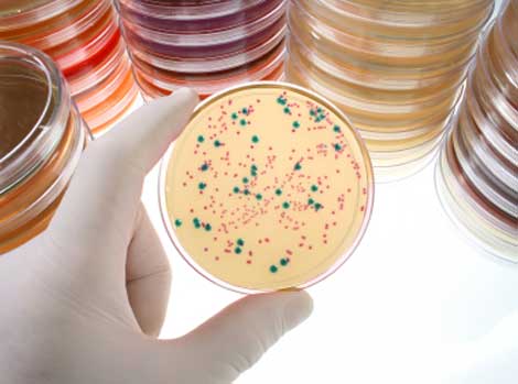
Agar plate containing colonies important for research, blue and white.
Agar plate containing colonies important for research.

Ah, the wonders and frustrations of cloning. We’ve all been there. After careful planning, you have created the cloned plasmid containing your DNA sequence of interest, transformed it into bacterial cells and carefully spread those cells on a plate to grow. Now you stand at your bench gazing down at your master piece: a plate full of tiny bacterial colonies. Somewhere inside those cells is your DNA sequence, happily replicating with its plasmid host. But wait – logic tells you that not ALL of those colonies can contain your plasmid.  There must be hundreds of colonies. Which ones have your plasmid? You begin to panic. Visions of yourself old and grey and still screening colonies flash through your mind. At the next bench, your lab-mate is cheerfully selecting colonies to screen. Although there are hundreds of colonies on her plate as well, some are white and some are blue. She is only picking the white colonies. What does she know that you don’t?

Continue reading “Selecting the Right Colony: The Answer is There in Blue and White”

In Vitro Transcription: Common Causes of Reaction Failure

FemaleWhiteLab-AAES001042, In Vitro Transcription

A widely used molecular biology technique, in vitro transcription uses bacteriophage DNA-dependent RNA polymerases to synthesize template-directed RNA molecules. Enzymes like bacteriophage SP6, T3 and T7 RNA polymerases are used to produce synthetic RNA transcripts, which can be used as hybridization probes, as templates for in vitro translation applications, or in structural studies (X-ray crystallography and NMR). Synthesized RNA transcripts are also used for studying cellular RNA functionality in processes such as splicing, RNA processing, intracellular transport, viral infectivity and translation.

Problems in the transcription reaction can result in complete failure (i.e., no transcript generated) or in transcripts that are the incorrect size (i.e., shorter or longer than expected). Below is a discussion of the most common causes of in vitro transcription problems.

Continue reading “In Vitro Transcription: Common Causes of Reaction Failure”

For the Birds: Knitting Nests for Baby Birds Might Just Help Your Health To

It seems that spring has finally come to Southern Wisconsin. The snow has melted. Most days it is warm enough you can go outside without a parka, hat and mittens. The tree buds are starting to swell. And that traditional oracle of spring, the American robin (Turdus migratorius), has been spotted in trees and yards—along with its less friendly cousin, the red winged black bird (Agelaius phoeniceus).

While spring brings the return of migratory birds, it also brings an increase in the number of rescued baby birds flooding into local wildlife rescues and humane societies. When the babies come to these centers, they need a warm, soft, breathable and washable home that resembles the nest they were hatched in.

It turns out that knitted or crocheted nests are a perfect solution. The nests aren’t just used for baby birds; baby rabbits, squirrels, bats, ferrets and racoons are just a few additional animals that benefit. And the best part is, you could be improving your own health while you create those cozy nests. Continue reading “For the Birds: Knitting Nests for Baby Birds Might Just Help Your Health To”

Cloning Modified Blunt-ended DNA Fragments into T-Vectors

Tailing blunt-ended DNA fragments with TaqDNA Polymerase allows efficient cloning of these fragments into T-Vectors such as the pGEM®-T Vectors. This method also eliminates some of the requirements of conventional blunt-end cloning — Fewer steps, who can argue with that?

Blue/White colony screening helps you pick only the colonies that have your insert.

Continue reading “Cloning Modified Blunt-ended DNA Fragments into T-Vectors”