In 2000 measles was officially declared eliminated in the United States (1), meaning there had been no disease transmission for over 12 months. Unfortunately, recent years have shown us it was not gone for good. So far in 2025 there have been 6 outbreaks and 607 cases. Five hundred and sixty-seven of these cases (93%) are associated with an outbreak; seventy-four (12%) cases have resulted in hospitalization, and there has been one confirmed death, with another death under investigation (as of April 3, 2025; 2). For comparison, there were two hundred and eighty-five total cases in 2024; one hundred and ninety-eight (69%) were associated with outbreaks; one hundred and fourteen (40%) cases resulted in hospitalization. There were no deaths (2).
Help in Limiting a Dangerous Childhood Disease
Before the development of a vaccine in the 1960s, measles was practically a childhood rite of passage. This common childhood disease is not without teeth however. One out of every 20 children with measles develops pneumonia, 1 out of every 1,000 develops encephalitis (swelling of the brain), and 1 to 3 of every 1,000 dies from respiratory and neurological complications (3). In the years before a vaccine was available, it is estimated that there were between 3.5 and 5 million measles cases per year. (4). The first measles vaccine was licensed in the U.S. by John Enders in 1963, and not surprisingly, after the measles vaccine became widely used, the number of cases of measles plummeted. By 1970, there were under 1,000 cases (2).
Decreased Childhood Mortality from Other Infectious Diseases—An Unexpected Benefit
Surprisingly, with the disappearance of this childhood disease the number of childhood deaths from all infectious diseases dropped dramatically. As vaccination programs were instituted in England and parts of Europe, the same phenomenon was observed. Reduction or elimination of measles-related illness and death alone can’t explain the size of the decrease in childhood mortality. Although measles infection is associated with suppression of the immune system that will make the host vulnerable to other infections, these side effects were assumed to be short lived. In reality, the drop in mortality from infectious diseases following vaccination for measles lasted for years, not months (5).
Three of the most common metrics in drug discover are Kd, IC50 and EC50. At first glance it can seem that they measure the same thing, but they don’t. Kd measures how tightly a molecule or compound binds to its target. IC50 measures inhibition of a function and conversely, EC50 measures activation or induction of a response. Confusing these values can lead to misinterpretation of assay results and costly rework. Let’s take a closer look at each one.
As climate change accelerates, understanding how crops survive environmental stress isn’t just an academic question—it’s a critical challenge for global food security. Tomatoes (Solanum lycopersicum), a staple crop worldwide, face increasing threats from drought, salinity, and extreme temperatures. But how do these plants adapt at the molecular level?
A recent study published in Scientific Reports investigated the evolutionary history, genomic diversity, and functional roles of protein phosphatase 2C (PP2C) genes in tomatoes (1). Instead of merely cataloging these genes, the researchers analyzed how PP2C gene expression changes under environmental stress. This information could help inform us about crop improvement strategies.
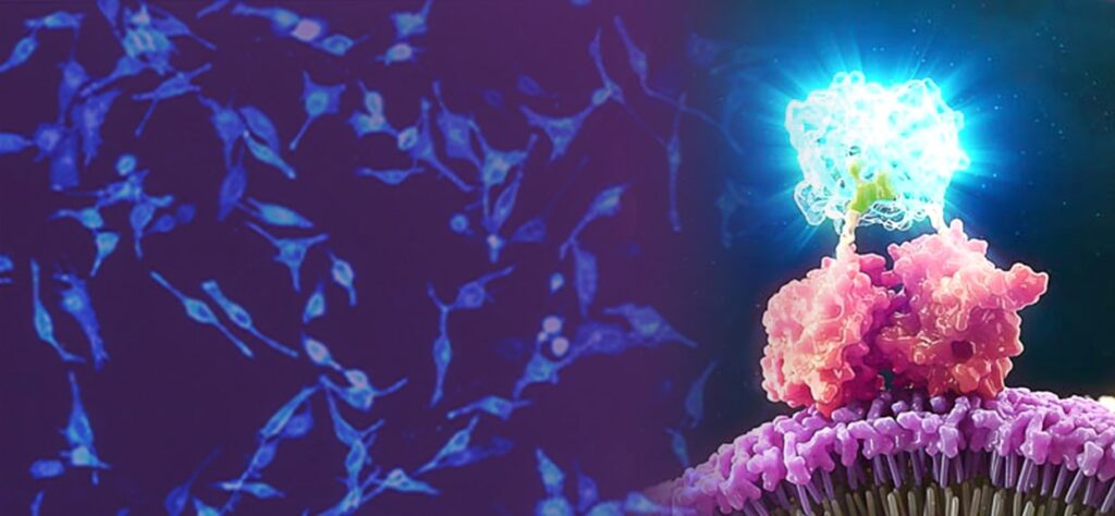
Luminescent live-cell assays are powerful tools for cellular biology research. They offer both qualitative and quantitative insights into processes such as gene expression, cell viability, metabolic activity, protein and small molecule interactions, and targeted protein degradation. But what if you could go beyond the numbers and actually see what is happening in your cells? With luminescent imaging, you have the opportunity to uncover more dynamic data by visualizing what happens with your cells in real time.
Why Luminescent Imaging?
Bioluminescent reporters such as NanoLuc® Luciferase reporters are well-suited for use in bioluminescent imaging studies. The extreme brightness means that exposure times can be reduced, compared to the time required for other luminescent reporter proteins. Its small size also makes it less likely to perturb the normal biology or functionality.
Another benefit of bioluminescence for imaging is the inherent stability and sustainability of the bioluminescent signal, which does not require external excitation like fluorescent tags. This allows direct visualization of protein dynamics in living cells without the need for repeated sample excitation. The lack of external excitation also reduces the risk of phototoxicity and photobleaching, common issues that can adversely affect cell viability and signal integrity over time.
Applications Across Cellular Research
Luminescent imaging complements traditional luminescence assays by adding spatial and temporal dimensions. With luminescent live-cell imaging, researchers can visualize NanoLuc® Luciferase assays to gain a deeper understanding of the real-time cellular processes occurring in each experiment. Applications include:
Determining which cells provide signal
Analyzing mixed cell populations
Identifying rare events
Monitoring protein:protein interactions
Identifying protein localization and translocation
Tracking protein degradation and stability over time
Selectively targeting proteins for removal from the cell—instead of inhibiting protein activity—is a newer approach with therapeutic potential. In this method, the protein is targeted for degradation using the cell’s natural ubiquitin proteasome system (UPS). The degradation process is initiated by compounds such as molecular glues and proteolysis targeting chimeras (PROTACs) linking the target protein to an E3 ligase. Once this linkage occurs, the cell’s UPS does the rest.
Luminescent substrates with increased signal stability, such as the Nano-Glo® Extended Live Cell Substrate, enables researchers to image targeted protein degradation in their cells in real time. In the example shown below, Nano-Glo® Vivazine™ Live Cell Substrate was used to image degradation of the GSPT1 protein by the CC-885 degrader over 5 hours.
Targeted protein degradation over time. HEK293 cells expressing endogenous HiBiT-tagged GSPT1 and stably expressing LgBiT were treated with CC-885 degrader or DMSO control treatment. Assayed with Nano-Glo® Vivazine™ Live Cell Substrate and imaged over 5 hours using GloMax® Galaxy Bioluminescence Imager.
Combining Luminescent and Fluorescent Imaging to Detect Protein:Small Molecule Interactions
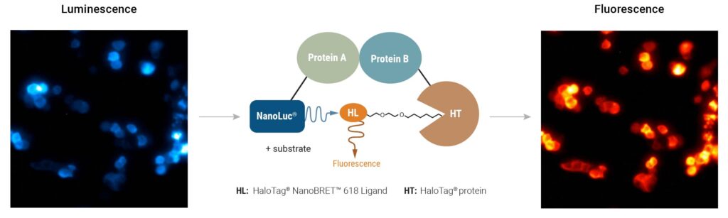
Using bioluminescence resonance energy transfer (BRET)-based assays such as NanoBRET® assays allows you to detect protein:protein interactions by measuring energy transfer from a bioluminescent protein donor to a fluorescent protein acceptor. These assays can be used to monitor changes in protein interactions over time, making them a useful tool for small-molecule screening.
The schematic below illustrates how the NanoBRET® NanoGlo® Detection Systems can be used to visualize target engagement. The cells on the left are expressing a NanoLuc® fusion protein, resulting in a luminescent signal. Adding a fluorescent small tracer (center) results in energy transfer and a fluorescent signal (right). Using an imaging platform that has luminescence and fluorescence imaging capabilities will let you see this energy transfer in action.
Detecting protein:small molecule interactions with NanoBRET® NanoGlo® Detection Systems. HCT116 cells expressing a PRMT5–NanoLuc® fusion were supplemented with a fluorescent small molecule tracer (center panel). Before tracer addition, luminescent signal indicates energy is present on the donor protein (left; 3-minute exposures for 15 minutes). Binding of fluorescent tracer results in energy transfer and fluorescent signal (right; 3-minute exposures for 60 minutes). Images were captured on the GloMax® Galaxy Bioluminescence Imager.
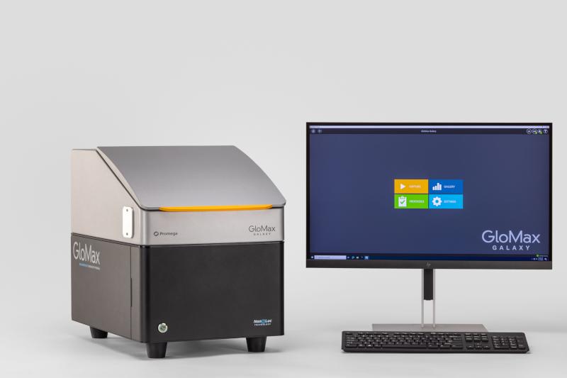
Bringing the Power of Luminescent Imaging to Your Lab
Having the right tools is critical to unlocking the full potential of bioluminescence imaging. The GloMax® Galaxy Bioluminescence Imager is uniquely positioned to offer researchers the power of imaging in an accessible, benchtop instrument. The Galaxy is a fully equipped microscope that can visualize output from NanoLuc® Technologies and offers luminescence, fluorescence and brightfield imaging capabilities. By offering a user-friendly platform for live-cell luminescent imaging, the GloMax® Galaxy empowers researchers to enrich their understanding of functional and dynamic cellular events across a cell population.
Conclusion
Luminescent imaging can enrich what we learn from live-cell assays and offers an unprecedented view into the dynamics of cellular processes. From monitoring drug responses to visualizing protein interactions, this technology delivers insights that go beyond the capabilities of traditional assays.
Whether you’re studying cancer biology, drug development or cellular signaling, luminescent imaging can help you uncover what’s hidden in your data and see your research in a whole new light.
GloMax® Galaxy Luminescent Imager, NanoBRET® Nano-Glo® Detection Systems and Nano-Glo® Vivazine live Cell Substrate are for Research Use Only. Not for Use in Diagnostic Procedures.
Southern sea otters (Enhydra lutris nereis), endangered marine mammals along California’s coastlines, are facing an unexpected threat. The menace comes not from pollution, habitat loss or natural predators, but from a microscopic enemy—Toxoplasma gondii (T. gondii). This protozoan parasite, typically associated with domestic cats, has found its way into marine ecosystems with sometimes deadly consequences for sea otters. Recently, scientists identified transmission of virulent, atypical strains of T. gondii from terrestrial felids to sea otters along the southern California coast, with lethal consequences (1).
Understanding T. gondii and Its Hosts
T. gondii is a versatile parasite that can infect nearly all warm-blooded animals, including humans and marine mammals. However, the T. gondii lifecycle depends upon felids (e.g., domestic cats and their wild relatives) who serve as definitive hosts. It is in their intestines that the parasite completes its sexual reproductive stage. The resulting oocysts are excreted in the animals’ feces. T. gondii oocysts exhibit remarkable resilience, surviving in soil, freshwater and seawater for extended periods. They are even resistant to standard wastewater treatment processes, which means oocysts in cat waste disposed of by flushing will pass through the treatment plant and be discharged into the environment. (2,3).
Oocysts can also be washed from soil contaminated with cat waste and carried via storm drains and rivers into the ocean, dispersing them into coastal waters. Once there, the oocysts settle on kelp or in sediments where they can be picked up by marine invertebrates like snails, mussels and clams. Marine mammals such as sea otters become infected when they consume these contaminated invertebrates. Otters can also ingest oocysts during grooming sessions (1,3).
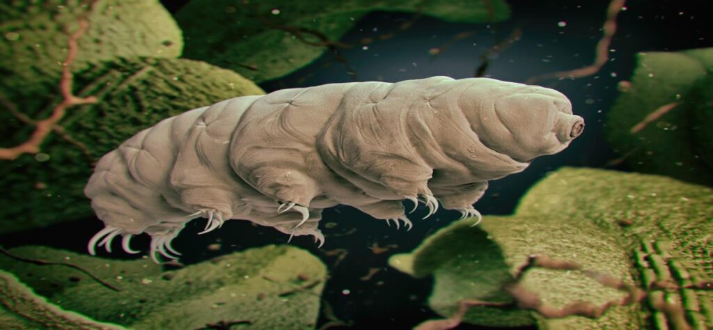
Some of our most advanced medicines today rely on components derived from living organisms. These therapeutics, called biologics, include things like vaccines, blood products like Human Blood Clotting Factor VIII (FVIII), antibodies and stem cells. Biologics are incredibly temperature sensitive, which means they need to be kept cold during production, transport and storage, a process collectively called the cold chain. The stringent transport and storage temperature requirements for biologics create a barrier to accessing these lifesaving options; particularly for those in remote or underdeveloped regions, where maintaining a cold chain is logistically difficult and costly.
But what if we could break the cold chain? Inspired by one of the most resilient creatures on Earth – the tardigrade – scientists at the University of Wyoming are exploring ways to do just that.
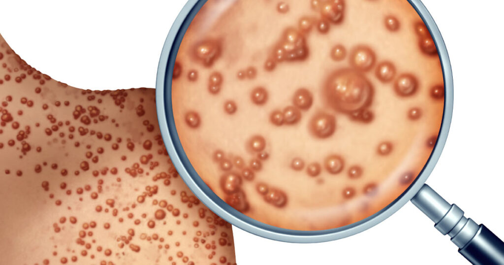
Mpox (formerly known as Monkeypox; 1) has been making the news lately. The declaration by the WHO Director-General naming mpox a public health emergency of international concern (PHEIC; 2) has a lot of people wondering what it is, how it spreads and how concerned they should be. Understandably, we are all a little jumpy when we start hearing about a new viral disease, but the virus that causes mpox (monkeypox virus) isn’t new.
A member of the Poxviridae family, the monkeypox virus is closely related to the variola virus that causes smallpox; however, monkeypox causes milder symptoms and is less fatal (1). While the virus gained its unfortunate name from its discovery in monkeys in 1958 (3), the original source of the disease remains unknown. The virus exists in a wide range of mammals including rodents, anteaters, hedgehogs, prairie dogs, squirrels and shrews (4) and can spread to humans through close contact with an infected individual or animal. Symptoms can include fever, headache, muscle and back pain, swollen lymph nodes, chills and exhaustion (3). The most distinguishing symptom is the blister-like rash.
It sounds like the script for a Hollywood movie. The story, first appearing in 2001, begins with a purported civil war legend from the Battle of Shiloh. The legend said that the wounds of some soldiers glowed (faintly) in the dark. Soldiers with these glowing wounds were more apt to survive, giving the phenomenon the name “Angels Glow”. The story ends with two curious teenagers solving the mystery using their science fair project. They identify infection by the bioluminescent bacteria Photorhabdus luminescens (formerly Xenorhabdus luminescens) as the likely cause of the glowing wounds. P. luminescens produces bacteriocins (antimicrobial peptides), which the teenagers attribute to helping keep other infections at bay, resulting in the improved survival rate for the soldiers whose wounds glowed.
The teenagers win. The mystery is solved. The credits roll.
Except life (and science) is rarely as simple as a summer block buster.
The Battle of Shiloh took place in Hardin County Tennessee on April 6th and 7th, 1862.
For the first time since Thomas Jefferson was president, broods of 13- and 17-year periodical cicadas are emerging from the ground at the same time. The fate that awaits some of these periodic cicadas—a fungal infection that hijacks their behavior and destroys their genitalia — sounds like the script of a bad zombie horror film. The culprit (or villain) is the entomopathogenic fungus Massospora cicadina.
While most entomopathogens kill their host before releasing their infectious spores, M. cicadina is one of the few species that increase spore dispersal by hijacking their host’s behavior and keeping them alive while sporulating (1). The manner it uses to do this is both gruesome and fascinating. If you can stomach some details of insect sex and dismemberment, read on.
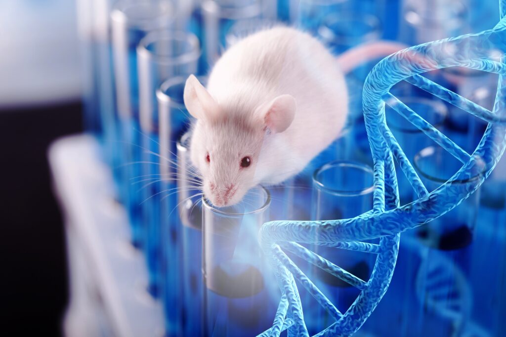
Amyotrophic lateral sclerosis (ALS) and frontotemporal dementia (FTD) are fatal and rapidly progress as neurodegenerative diseases. While inherited mutations can cause both conditions, they mostly appear sporadically in individuals without a known family history. Despite affecting different neurons, both diseases share a common hallmark: the pathogenic buildup of abnormal nuclear TAR-binding protein 43 (TDP-43) in the cytoplasm of affected motor neuron cells. Current theories propose that this cytoplasmic re-localization triggers toxic phosphorylation and fragmentation of TDP-43. Concurrently, a decrease of TDP-43 in the nucleus diminishes TDP-43-related physiological nuclear functions, contributing to the diseases’ progression (1).
Although this cytoplasmic accumulation of TDP-43 plays a significant role in the pathogenesis of ALS and FTD, the cellular mechanisms involved in the re-localization of TDP-43 to the cytoplasm is not known (2). A team of Australian neuroscientists led by Dr. Lars Ittner believe that they have found part of the answer for sporadic forms of the diseases. They identified novel interactions between pathogenic or dysfunctional forms of TDP-43 and the 14.3.3ɵ isoform of the cytoplasmic protein 14-3-3. By targeting this interaction with an AAV-based gene therapy vector, they were able to block and even partially reverse neurodegeneration in ALS/FTD mouse models.
XWe use cookies and similar technologies to make our website work, run analytics, improve our website, and show you personalized content and advertising. Some of these cookies are essential for our website to work. For others, we won’t set them unless you accept them. To learn more about our approach to Privacy we invite you to Read More
By clicking “Accept All”, you consent to the use of ALL the cookies. However you may visit Cookie Settings to provide a controlled consent.
We use cookies and similar technologies to make our website work, run analytics, improve our website, and show you personalized content and advertising. Some of these cookies are essential for our website to work. For others, we won’t set them unless you accept them. To find out more about cookies and how to manage cookies, read our Cookie Policy.
If you are located in the EEA, the United Kingdom, or Switzerland, you can change your settings at any time by clicking Manage Cookie Consent in the footer of our website.
Necessary cookies are absolutely essential for the website to function properly. These cookies ensure basic functionalities and security features of the website, anonymously.
Cookie
Duration
Description
cookielawinfo-checbox-analytics
11 months
This cookie is set by GDPR Cookie Consent plugin. The cookie is used to store the user consent for the cookies in the category "Analytics".
cookielawinfo-checbox-functional
11 months
The cookie is set by GDPR cookie consent to record the user consent for the cookies in the category "Functional".
cookielawinfo-checbox-others
11 months
This cookie is set by GDPR Cookie Consent plugin. The cookie is used to store the user consent for the cookies in the category "Other.
cookielawinfo-checkbox-advertisement
1 year
The cookie is set by GDPR cookie consent to record the user consent for the cookies in the category "Advertisement".
cookielawinfo-checkbox-necessary
11 months
This cookie is set by GDPR Cookie Consent plugin. The cookies is used to store the user consent for the cookies in the category "Necessary".
cookielawinfo-checkbox-performance
11 months
This cookie is set by GDPR Cookie Consent plugin. The cookie is used to store the user consent for the cookies in the category "Performance".
gdpr_status
6 months 2 days
This cookie is set by the provider Media.net. This cookie is used to check the status whether the user has accepted the cookie consent box. It also helps in not showing the cookie consent box upon re-entry to the website.
lang
This cookie is used to store the language preferences of a user to serve up content in that stored language the next time user visit the website.
viewed_cookie_policy
11 months
The cookie is set by the GDPR Cookie Consent plugin and is used to store whether or not user has consented to the use of cookies. It does not store any personal data.
Analytical cookies are used to understand how visitors interact with the website. These cookies help provide information on metrics the number of visitors, bounce rate, traffic source, etc.
Cookie
Duration
Description
SC_ANALYTICS_GLOBAL_COOKIE
10 years
This cookie is associated with Sitecore content and personalization. This cookie is used to identify the repeat visit from a single user. Sitecore will send a persistent session cookie to the web client.
vuid
2 years
This domain of this cookie is owned by Vimeo. This cookie is used by vimeo to collect tracking information. It sets a unique ID to embed videos to the website.
WMF-Last-Access
1 month 18 hours 24 minutes
This cookie is used to calculate unique devices accessing the website.
_ga
2 years
This cookie is installed by Google Analytics. The cookie is used to calculate visitor, session, campaign data and keep track of site usage for the site's analytics report. The cookies store information anonymously and assign a randomly generated number to identify unique visitors.
_gid
1 day
This cookie is installed by Google Analytics. The cookie is used to store information of how visitors use a website and helps in creating an analytics report of how the website is doing. The data collected including the number visitors, the source where they have come from, and the pages visted in an anonymous form.
Advertisement cookies are used to provide visitors with relevant ads and marketing campaigns. These cookies track visitors across websites and collect information to provide customized ads.
Cookie
Duration
Description
IDE
1 year 24 days
Used by Google DoubleClick and stores information about how the user uses the website and any other advertisement before visiting the website. This is used to present users with ads that are relevant to them according to the user profile.
test_cookie
15 minutes
This cookie is set by doubleclick.net. The purpose of the cookie is to determine if the user's browser supports cookies.
VISITOR_INFO1_LIVE
5 months 27 days
This cookie is set by Youtube. Used to track the information of the embedded YouTube videos on a website.
Performance cookies are used to understand and analyze the key performance indexes of the website which helps in delivering a better user experience for the visitors.
Cookie
Duration
Description
YSC
session
This cookies is set by Youtube and is used to track the views of embedded videos.
_gat_UA-62336821-1
1 minute
This is a pattern type cookie set by Google Analytics, where the pattern element on the name contains the unique identity number of the account or website it relates to. It appears to be a variation of the _gat cookie which is used to limit the amount of data recorded by Google on high traffic volume websites.