Understanding how a compound or drug affects cellular pathways often requires measuring kinetic changes over an extended period of time—from several hours to days. Live-cell kinetic cell-based assays that measure cell viability, cytotoxicity, apoptosis and other cellular pathways are great for collecting real-time data. You don’t necessarily need expensive equipment to run these types of assays. In the videos below, Dr. Sarah Mahan, a research scientist at Promega, demonstrates how you can easily get great 24-hour or multi-day kinetic data using a GloMax® Microplate Reader.
Continue reading “How to Get Real-Time Kinetic Data With GloMax® Microplate Readers”Author: Johanna Lee
How A New Wastewater Surveillance Method Predicted COVID-19 Cases in Italy
Wastewater-based epidemiology (WBE), or sewershed surveillance, is the analysis of wastewater to identify the presence of biologicals or chemicals for the purpose of monitoring public health. In the past, WBE has been used to detect the presence of pharmaceutical or industrial waste, drugs and viruses. Now, it is seen as a valuable tool to monitor COVID-19 outbreaks.
Continue reading “How A New Wastewater Surveillance Method Predicted COVID-19 Cases in Italy”Rare Human Antibody Could Lead to A Universal Coronavirus Vaccine
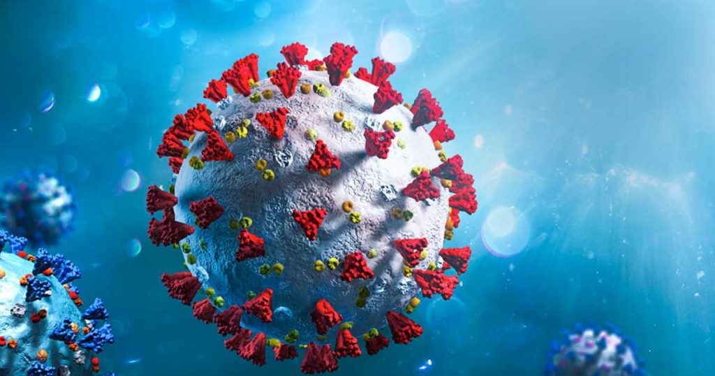
As we’ve all learned from the current global COVID-19 pandemic, coronaviruses are generally bad news. Among the four genera of coronaviruses, the betacoronavirus genus has been especially notorious. In the past 20 years, three highly pathogenic betacoronaviruses, MERS-CoV, SARS-CoV and SARS-CoV-2 have resulted in serious pandemics. All three of these viruses originated from bats, highlighting the continued risk of coronavirus transmission from animals to humans.
Typically, when a new viral threat emerges, researchers scramble to develop drugs or vaccines after the virus has already gotten out of control. However, developing a vaccine for each new virus is a slow and painstaking process, and many lives will be lost before a vaccine is distributed. But what if we had one, universal coronavirus vaccine that could neutralize not only all existing betacoronaviruses, but any new variants that emerge in the future?
Continue reading “Rare Human Antibody Could Lead to A Universal Coronavirus Vaccine”New Therapy for Brain Tumors: High-Pressure Oxygen Rewires Glucose Metabolism in Glioblastoma
Glioblastoma (GBM) is an aggressive type of brain tumor, and one of the deadliest cancers. GBM is often treated with surgery, radiation and chemotherapy, but even if the initial treatment is successful, a majority of patients relapse within months. One reason why GBM is so difficult to treat is the hypoxic (low-oxygen) tumor environment. It is known that hypoxic cells are resistant to radiotherapy; the greater the number of tumor stem cells in a hypoxic environment, the less efficient radiotherapy is at controlling tumor growth.
A new therapeutic approach aims to remove the hypoxic environment in GBM by administering pure oxygen to patients at high pressure, known as “hyperbaric oxygen (HBO) therapy”. Previous studies have shown that HBO improves the efficacy of radiotherapy in GBM patients. However, the therapeutic mechanism of HBO was largely unknown. That is, until now.
Dr. Anna Tesei, the Head of Radiobiomics and Drug Discovery at the Biosciences Laboratory of IRST-IRCCS in Italy, recently published a study on the mechanism in which HBOT affects GBM tumor cells and the tumor environment. “The main purpose of our study was to provide a preclinical rationale for the use of hyperbaric oxygen in association with radiotherapy for the treatment of GBM,” she says.
From Drug Use to Viral Outbreaks, How Monitoring Sewage Can Save Lives
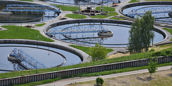
Most of us, after we flush the toilet, don’t think twice about our body waste. To us, it’s garbage. To epidemiologists, however, wastewater can provide valuable information about public health and help save lives.
History of Wastewater-Based Epidemiology
Wastewater-based epidemiology (WBE) is the analysis of wastewater to monitor public health. The term first emerged in 2001, when a study proposed the idea of analyzing wastewater in sewage-treatment facilities to determine the collective usage of illegal drugs within a community. At the time, this idea to bridge environmental and social sciences seemed radical, but there were clear advantages. Monitoring wastewater is a nonintrusive and relatively inexpensive way to obtain real-time data that accurately reflects community-wide drug usage while ensuring the anonymity of individuals.
Continue reading “From Drug Use to Viral Outbreaks, How Monitoring Sewage Can Save Lives”Oncolytic Viruses: Models and Assays for Developing Viruses That Can Kill Cancer

When we think of viruses, we often think of diseases, pandemics and death. Our impression of viruses is that they are “bad”. But viruses could also be a possible cure for the deadliest disease in modern history: cancer. The therapeutic effects of “good” cancer-killing oncolytic viruses have been documented over a century ago. Records from as early as 1904 described a 42-year old woman with acute leukemia who experienced temporary remission after an influenza infection. Other early reports showed spontaneous remission of Hodgkin lymphoma and Burkitt’s lymphoma after natural infections with the measles virus.
Despite the long history, oncolytic viruses have only recently gained momentum in the scientific community. Dr. Aldo Pourchet, CSO and co-founder of Omios Biologics—a biotech startup in the San Francisco Bay area—is determined to harness the power of oncolytic viruses to develop a new generation of cancer immunotherapy.
How Oncolytic Viruses Work
“One thing that we know for sure is that you need the immune system to fight the cancer,” says Pourchet. “You need to recruit the immune system, and probably the best thing we know for recruiting the immune system is viruses. Our immune system evolved to detect them immediately. That’s why we are still on Earth. It’s because we have been able to fight deadly viruses.”
Continue reading “Oncolytic Viruses: Models and Assays for Developing Viruses That Can Kill Cancer”Understanding Inflammation: A Faster, Easier Way to Detect Cytokines in Cells
Inflammation, a process that was meant to defend our body from infection, has been found to contribute to a wide range of diseases, such as chronic inflammation, neurodegenerative disorders—and more recently, COVID-19. The development of new tools and methods to measure inflammation is crucial to help researchers understand these diseases.
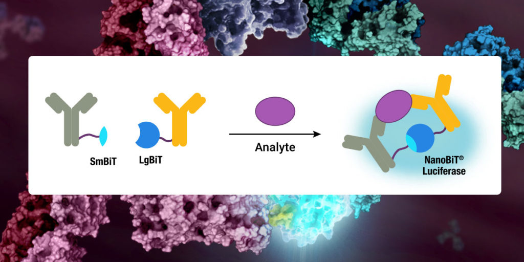
Cytokines—small signaling molecules that regulate inflammation and immunity—have recently become the focus of inflammation research due to their role in causing severe COVID-19 symptoms. In these severe cases, the patient’s immune system responds to the infection with uncontrolled cytokine release and immune cell activation, called the “cytokine storm”. Although the cytokine storm can be treated using established drugs, more research is needed to understand what causes this severe immune response and why only some patients develop it.
Continue reading “Understanding Inflammation: A Faster, Easier Way to Detect Cytokines in Cells”Comparing Methods for Detecting SARS-CoV-2 in Wastewater
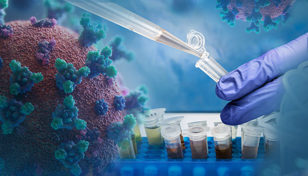
Wastewater surveillance of SARS-CoV-2 is an increasingly common method for monitoring the spread of COVID-19 within a community. As researchers and public health officials around the world are working together to set up wastewater surveillance systems, there is an urgent need to establish standard SARS-CoV-2 detection methods.
A key leader in this new field is Dr. Gloria Sanchez. She is a tenured scientist at the Institute of Agrochemistry and Food Technology, a center within the Spanish National Research Council. Before the COVID-19 pandemic, her team focused on detecting human enteric viruses in food and water. But soon, detecting SARS-CoV-2 in wastewater became their main focus.
Continue reading “Comparing Methods for Detecting SARS-CoV-2 in Wastewater”Monochromator vs Filter-Based Plate Reader: Which is Better?
When it comes to purchasing a microplate reader for fluorescence detection, the most common question is whether to choose a monochromator-based reader or filter-based reader. In this blog, we’ll discuss how both types of plate readers work and factors to consider when determining the best plate reader for your need.
How do monochromator-based plate readers work?
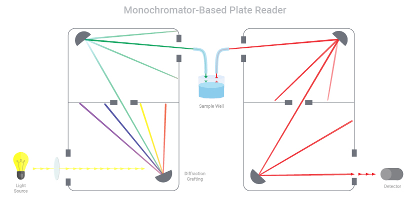
Monochromators work by taking a light source and splitting the light to focus a particular wavelength on the sample. During excitation, the light passes through a narrow slit, directed by a series of mirrors and diffraction grating and then passes through a second narrow slit prior to reaching the sample. This ensures the desired wavelength is selected to excite the fluorophore. Once the fluorophore is excited, it emits light at a different, longer wavelength. This emission light is captured by another series of mirrors, grating and slits to limit the emission to a desired wavelength, which then enters a detector for signal readout.
A SARS-CoV-2 NanoLuc® Reporter Virus for Rapid Screening of Antivirals

Before the COVID-19 global pandemic began, Dr. Xuping Xie, Assistant Professor of the University of Texas Medical Branch at Galveston, TX has been studying viruses, such as Dengue and Zika, for more than 10 years. Once the pandemic hit in early 2020, he was prepared to join the fight against the virus. “There was an urgent need to know: Is there a quicker way to develop therapeutics or antibodies to target SARS-CoV-2?” says Dr. Xie. “That’s why we immediately launched our SARS-CoV-2 project.”
His goal was to create an assay that could 1) screen for antiviral drugs and 2) quickly measure neutralizing antibody levels. The assay could be used to determine the immune status of previously infected individuals and to evaluate various vaccines under development. To achieve this, he wanted to create a reporter virus that is genetically stable and replicates similarly to the wild-type virus in cell culture.
Continue reading “A SARS-CoV-2 NanoLuc® Reporter Virus for Rapid Screening of Antivirals”