 Cell free protein expression can be utilized for the analysis of: protein/protein interactions, protein nucleic acid interactions, analysis of post translational modifications and many other applications. The majority of these references are based on the characterization of mammalian proteins.
However there are several references using TNT-based systems (either rabbit reticulocyte lysate or wheat germ based) for the analysis of proteins from plants, examples include: Continue reading “Cell-Free Protein Expression: Characterization of Plant Proteins”
Author: Gary Kobs
Use of Cell-Free Protein Expression for Epigenetics-Related Applications
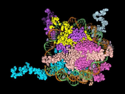
 Epigenetics is the study of the processes involved in the genetic development of an organism, especially the activation and deactivation of genes. One way that genes are regulated is through the remodeling of chromatin. Chromatin is the complex of DNA and the histone proteins with which it associates. The conformation of chromatin is profoundly influenced by the post-translational modification of the histone proteins. These modifications include acetylation, methylation, ubiquitylation, phosphorylation and sumolyation. The following references illustrate the use of cell-free expression to characterize this process.
Shao, Y. et al. (2010) Nucl. Acid. Res. 38, 2813–24.
Carbonic anhydrase IX (CAIX) plays an important role in the growth and survival of tumor cells.The MORC proteins contain a CW-type zinc finger domain and are predicted to have the function of regulating transcription, but no MORC2 target genes have been identified. CAIX mRNA to be down-regulated 8-fold when MORC2 was overexpressed. Moreover, MORC2 decreased the acetylation level of histone H3 at the CAIX promoter. Among the six HDACs tested, histone deacetylase 4 (HDAC4) had a much more prominent effect on CAIX repression. Assays showed that MORC2 and HDAC4 were assembled on the same region of the CAIX promoter. Interaction between MORC2 and HDAC 4 were confirmed by using cell free expression of MORC2 and GST-HDAC (GST pull-downs). Cell-free expression was also used to express MORC2 proteins to determine through gel shifts the binding location on the CAIX promoter region (gel shift experiments)
Denis, H. et al. (2009) Mol. Cell. Biol. 29, 4982–93.
The recent identification of enzymes that antagonize or remove histone methylation offers new opportunities to appreciate histone methylation plasticity in the regulation of epigenetic pathways. PAD4 was the first enzyme shown to antagonize histone methylation. Very little is known as to how PADI4 silences gene expression. Through the use of cell-free expression to express both PAD4 and HDAC1 proteins and E. coli expression of GST fusions of PAD4 and HDAC1, pulldown experiments confirmed by in vivo experiments that PADI4 associates with the histone deacetylase 1 (HDAC1), and the corresponding activities, associate cyclically and coordinately with the pS2 promoter during repression phases.
Brackertz, M. et al. (2006) Nucl. Acid. Res. 34, 397-406.
The Mi-2/NuRD complex is a multi-subunit protein complex with enzymatic activities involving chromatin remodeling and histone deacetylation. The function of p66α and of p66β within the multiple subunits has not been addressed. GST-fused histone tails of H2A, H2B, H3 and H4 were expressed in E. coli used in an in vitro pull-down assay with radioactively labeled p66-constructs expressed using cell free systems. Deletions at the C terminus noted reduced binding of p66 where as deletions at the N terminus did not affect binding. Also observed was that acetylation of histone tails reduces the association with both p66-proteins in vitro.
Zhou, R. et al. (2009) Nucl. Acids. Res. 37, 5183–96.
Lymphoid specific helicase (Lsh) belongs to the family of SNF2/helicases. Disruption of Lsh leads to developmental growth retardation and premature aging in mice. However, the specific effect of Lsh on human cellular senescence remains unknown. In vivo results noted that Lsh requires histone deacetylase (HDAC) activity to repress p16INK4a. Moreover, overexpression of Lsh is correlated with deacetylation of histone H3 at the p16 promoter. In vitro pull-downs using cell free expression and GST fusions from E. coli were used to collaborate interactions between Lsh, histone deacetylase 1 (HDAC1) and HDAC2 observed in vivo.
Purification of biotinylated proteins
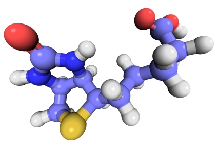
Biotinylation is an attractive approach for protein complex purification due to the very high affinity (Kd = 10–15 M) of avidin/streptavidin for biotinylated templates. With typical avidin or streptavidin, the biotin-binding affinity is so great that purification with these traditional media require denaturing conditions for elution,such as 8 M Guanidine•HCl at pH 1.5 or boiling in reducing SDS-PAGE sample loading buffer. To avoid these harsh conditions SoftLink™ Soft Release Avidin resin can be used. These particles consist of monomeric avidin coupled to a methylacrylate resin.
This resin provides the same specificity of binding to biotin afforded by tetrameric biotin, but enables the release of biotinlylated molecules under mild nondenaturing conditions (5mM biotin).
The following are recent references that used the SoftLink™ Resin for the noted application:
Kashwayama, Y. et al. (2010) Identification of a substrate-binding site in a peroxisomal beta-oxidation enzyme by photoaffinity labeling with a novel palmitoyl derivative. J. Biol. Chem. 285, 26315–25. (Purification of photoaffinity labeled proteins for subquenant binding/activity experiments)
Takahashi, M. et al. (2010) Tailor-made RNAi knockdown against triplet repeat disease-causing alleles. Proc. Natl. Acad. Sci. 107, 21731-36 (Innovated procedure using biotin labeled cDNAs for the identification of nucleotide variations)
Kress, D. et al. (2009) An asymmetric model for Na+-translocating glutaconyl-CoA decarboxylases. J. Biol.Chem. 284, 28401–9 (Purification of Clostridium biotin carrier proteins that play a role in decarboxylation)
Akahori, Y. et al. (2009) Characterization of neutralizing epitopes of varicella-zoster virus glycoprotein H. J. Virol. 83, 2020–4. (purification of double stranded cDNA fragments amplified by PCR with a biotin-tagged PCR primer)
Shonsey, E.M. et al. (2008) Inactivation of human liver bile acid CoA:amino acid N-acyltransferase by the electrophilic lipid, 4-hydroxynonenal. J. Lipid Res. 49, 282–94 (purification of recombinant protein expressed in E.coli containing C-terminal avidin tag)
Andachi, Y. et al. (2008) A novel biochemical method to identify target genes of individual microRNAs: identification of a new Caenorhabditis elegans let-7 target. RNA 14, 2440–51 (purification of double stranded cDNA fragments amplified by PCR with a biotin-tagged PCR primer)
For more information about SoftLink™ Soft Release Avidin Resin, please visit our website.
Cell Free Expression Application: Production of Soluble Protein for Structural Analysis
The TNT® SP6 High-Yield Protein Expression System uses a high-yield wheat germ extract supplemented with SP6 RNA polymerase and other components. Coupling transcriptional and translational activities eliminates the inconvenience of separate in vitro transcription and purification steps for the mRNA, while maintaining the high levels of protein expression. All that is required is the addition of DNA templates containing the SP6 promoter and the protein coding region for the protein of interest. Furthermore no specialized equipment is required for protein screening and production. The system enables the expression of approximately 100µg/ml of protein in batch reaction and 200–440µg/ml in dialysis reaction in 10–20 hours .
In a recent publication (Zhao, L. et.al. (2010) J. Struct. Genomics 11, 201–9), the Northeast Structural Genomics Consortium (www.nesg.org) in their quest to express 5,000 eukaryotic proteins, report that even with different cloning strategies they could only produce 26% of the proteins in a soluble form. To improve the efficiency of expressing soluble protein, they investigated the use of wheat germ cell free system as a alternative to E.coli.
In this publication 59 human constructs were expressed in both E.coli and the wheat germ cell free system. Only 30% of human proteins could be produced in a soluble form using E.coli -based expression. Some 70% could be produced using the TNT® SP6 High Yield Wheat Germ system.
To further demonstrate the utility of expressing proteins that were suitable for structural studies from a wheat germ-based system, two of the proteins were isotope enriched and analyzed successfully by 2D NMR.
Alternative Applications for Cell-Free Expression #3
Protein location: outer mitochondrial membrane (Yeast in vitro import assay)
Curado, S. et.al. (2010) Dis.Mod. Mech. 3, 486-95. PubMed ID 20483998.
Chemically mutagenized zebra fish were assayed for liver defects in their F3 progeny.This screen led to the identification of mutant called oliver. Oliver mutants have an o-shaped liver of a much deprived size due to the depletion of most of the hepatocytes. This mutation maps to the Tomm22gene which encodes a translocase of the outer membrane and thought to play an important role in protein import into mitochondria. Various Tomm22 mutants were expressed and used in a yeast in vitro import systemto determine if correct inserted into the yeast outer mitochondrial membrane.
Protein modification: hydroxylation
Serchov, T. et.al. (2010) J. Biol. Chem. 285, 21223-232. PubMed ID 20418372 .
Proline hydroxylation is also a vital component of hypoxia via hyposxia inducible factors. The cellular response to hypoxia involves the induction of the hypoxia-inducible factor considered to be the major transcription factor involved in gene regulation of hypoxia. This factor is hydroxylated by prolyl-hydroxase dolman proteins (PHDs). To investigate if a newly identified component of the hypoxia pathway (Elk3) is also hydroxylated, proteins were expressed +/- PHDs cofactors and protein mobility was measured via gel analysis.
Gene Experession: Programmed Ribosomal Frameshift
Kobayashi, Y. et.al. (2010) J. Biol. Chem. 285, 19776-784. PubMed ID 20427288.
Programmed -1 ribosomal frameshifting (PRF) is a distinctive mode of gene expression utilized by some viruses (HIV-1 for example). Recently a genome-wide screen demonstrated that down regulation of eukaryotic release factor (eRF1) inhibited HIV-1 replication. In order to characterize the dose dependent effect of eRF1, increasing amounts were expressed in the presence of dual luciferase reporter vectors harboring a HIV-1 PRF signal
Screening for Protein Activity Using Cell-Free Expression
The analysis of functional protein typically requires lengthy laborious cell based protein expression that can be complicated by the lack of stability or solubility of the purified protein. Cell free protein expression eliminates the requirement for cell culture thus providing quick access to the protein of interest (1).
The HaloTag® Technology provides efficient, covalent and oriented protein immobilization of the fusion protein to solid surfaces (2).
A recent publication demonstrated the feasibility of using cell free expression and the HaloTag technology to express and capture a fusion protein for the rapid screening of protein kinase activity (3). The catalytic subunit of human cAMP dependent protein kinase was expressed in a variety of cell free expression formats as a HaloTag fusion protein. The immobilized cPKA fusion protein was assayed directly on magnetic beads in the active form and was shown to be inhibited by known PKA inhibitory compounds.
Therefore this unique combination of protein expression and capture technologies can greatly facilitate the process of activity screening and characterization of potential inhibitors
- Zhao, K.Q. et al. (2007) Functional protein expression from a DNA based wheat germ cell-free system. J. Struc. Funct. Genomics. 8, 199-208.
- Los, G.V. and Wood, K. (2007) The HaloTag: A novel technology for cell imaging and protein analysis. Meth. Mol. Biol. 356, 195-208
- Leippe DM, Zhao KQ, Hsiao K, & Slater MR (2010). Cell-free expression of protein kinase a for rapid activity assays. Analytical chemistry insights, 5, 25-36 PMID: 20520741
Protease K Protection Assay: Cell Free Expression Application
Microsomal vesicles are used to study cotranslational and initial posttranslational processing of proteins. Processing events such as signal peptide cleavage, membrane insertion, translocation and core glycosylation can be examined by the transcription/translation of the appropriate DNA in the TNT® Lysate Systems when used with microsomal membranes.
The most general assay for translocation makes use of the protection afforded the translocated domain by the lipid bilayer of the microsomal membrane. In this assay protein domains are judged to be translocated if they are observed to be protected from exogenously added protease. To confirm that protection is due to the lipid bilayer addition of 0.1% non-ionic detergent (such as Triton® X-100) solubilizes the membrane and restores susceptibility to the protease.
Are you looking for proteases to use in your research?
Explore our portfolio of proteases today.
Many proteases have proven useful for monitoring translocation in this fashion including Protease K or Trypsin.
The following are examples illustrating this application:
- Minn, I. et al. (2009) SUN-1 and ZYG-12, mediators of centrosome-nucleus attachment, are a functional SUN/KASH pair in Caenorhabditis elegans. Mol. Biol. Cell. 20, 4586–95.
- Padhan, K. et al. (2007) Severe acute respiratory syndrome coronavirus Orf3a protein interacts with caveolin. J.Gen.Virol. 88, 3067–77.
- Tews, B.A. et al. (2007) The pestivirus glycoprotein Erns is anchored in plane in the membrane via an amphipathic helix. J.Biol.Chem. 282, 32730–41.
- Pidasheva, S. et al. (2005) Impaired cotranslational processing of the calcium-sensing receptor due to signal peptide missense mutations in familial hypocalciuric hypercalcemia. Hum. Mol. Gen. 14, 1679–90.
- Smith, D. et al. (2002) Exogenous peptides delivered by ricin require processing by signal peptidase for transporter associated with antigen processing-independent MHC class I-restricted presentation. J. Immun. 169, 99–107.
6X His Protein Pulldowns: An Alternative to GST
![]() Pull-down assays probe interactions between a protein of interest that is expressed as fusion protein (e.g.,
Pull-down assays probe interactions between a protein of interest that is expressed as fusion protein (e.g.,
(e.g., bait) and the potential interacting partners (prey).
In a pull-down assay one protein partner is expressed as a fusion protein (e.g., bait protein) in E. coli and then immobilized using an affinity ligand specific for the fusion tag. The immobilized
bait protein can then be incubated with the prey protein. The source of the prey protein depends on whether the experiment is designed to confirm an interaction or to identify new interactions. After a series of wash steps, the entire complex can be eluted from the affinity support using SDS-PAGE loading buffer or by competitive analyte elution, then evaluated by SDS-PAGE.
Successful interactions can be detected by Western blotting with specific antibodies to both the prey and bait proteins, or measurement of radioactivity from a [35S] prey protein. bait) and potential interacting partners (prey).
The most commonly used method to generate a bait protein is expression as a fusion protein contain a GST (glutathione-S transferase) tag in E. coli. This is followed by immobilization on particles that contain reduced glutathione, which binds to the GST tag of the fusion protein. The primary advantage of a GST tag is that it can increase the solubility of insoluble or semi-soluble proteins expressed in E. coli.
Among fusion tags, His-tag is the most widely used and has several advantages including: 1) It’s small in size, which renders it less immunogenically active, and often it does not need to be removed from the purified protein for downstream applications; 2) There are a large number of commercial vectors available for expressing His-tagged proteins; 3) The tag may be placed at either the N or C terminus; 4) The interaction of the His-tag does not depend on the tag structure, making it possible to purify otherwise insoluble proteins using denaturing conditions. Continue reading “6X His Protein Pulldowns: An Alternative to GST”
Use of Multiple Proteases for Improved Protein Digestion
One of the approaches to identify proteins by mass spectrometry includes the separation of proteins by gel electrophoresis or liquid chromatography. Subsequently the proteins are cleaved with sequence-specific endoproteases. Following digestion the generated peptides are investigated by determination of molecular masses or specific sequence. For protein identification the experimentally obtained masses/sequences are compared with theoretical masses/sequences compiled in various databases.
Trypsin is the favored enzyme for this application, for the following reasons: A) the peptides contain a basic residue (Arg or Lys) on the C terminus and thus are good candidates for collision induced activation (CAD) in tandem experiments (low charge states and high mass-to-charge ratios); B) it is relatively Inexpensive; and C) optimal digestion conditions have been well characterized.
An inherent limitation of trypsin is the size of the peptides that it generates. For most organisms > 50% of tryptic peptides are less than 6 amino acids, too small for mass spectrometry based sequencing.
Are you looking for proteases to use in your research?
Explore our portfolio of proteases today.
One recent publication examined the use of multiple proteases (trypsin, LysC, ArgC , AspN and GluC) in combination with either CAD or electron-based fragmentation (ETD) to improve protein identification (1). Their results indicated a significant improvement from a single protease digestion (trypsin), which yielded 27,822 unique peptides corresponding to 3313 proteins. In contrast using a combination of proteases with either CAD or ETD fragmentation methods yielded 92,095 unique peptides mapping to 3908 proteins.
Swaney DL, Wenger CD, & Coon JJ (2010). Value of using multiple proteases for large-scale mass spectrometry-based proteomics. Journal of proteome research, 9 (3), 1323-9 PMID: 20113005
Trypsin: Innovative Applications

Tryptic digestion of samples and subsequent analysis by mass spectrometry is a popular technique for the identification of proteins typically those related to interaction partners or biomarkers characterization. This powerful tool can also be used for less traditional experimental designs. Three examples are:
Continue reading “Trypsin: Innovative Applications”
