Convenient, Non-Radioactive Detection of Isoaspartate

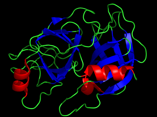
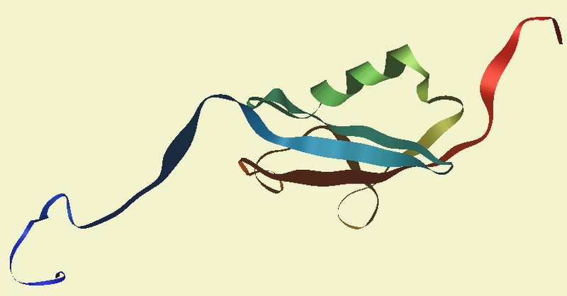
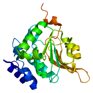
Structure of the PCMT1 protein. Based on PyMOL rendering of PDB 1i1n. Licensed under creative commons http://creativecommons.org/licenses/by-sa/3.0/deed.en

The ISOQUANT® Isoaspartate Detection Kit is intended for quantitative detection of isoaspartic acid residues in proteins and peptides, which can result from the gradual, nonenzymatic deamidation of asparagine or rearrangement of aspartic acid residues.

The ISOQUANT® Kit is designed to provide information regarding the global formation of isoaspartic acid residues at Asn and Asp sites, not at each site separately.

The deamidation of asparagine residues and rearrangement of aspartic acid residues is characterized by the formation of a succinimide intermediate that resolves to form a mixture of isoaspartic acid (typically 70–85%) and aspartic acid.
The rate and extent of isoaspartic acid formation can vary widely among proteins, depending on the amino acid sequence and size of the target protein. Deamidation of Asn residues has been observed most frequently at Asn-Gly and Asn-Ser sites within proteins.

The ISOQUANT® Isoaspartate Detection Kit uses the enzyme Protein Isoaspartyl ethyltransferase (PIMT) to specifically detect the presence of isoaspartic acid residues in a target protein. PIMT catalyzes the transfer of a methyl group from S-adenosyl-L-methionine (SAM) to isoaspartic acid. Spontaneous decomposition of this methylated intermediate results in the release of methanol and reformation of the succinimide.

References:

Wang, W. et al. (2012) Quantification and characterization of antibody deamidation by peptide mapping with mass spectrometry. Int. J. Mass. Spec. 312, 107–13.

Grappin, P. et al. (2011) New proteomic developments to analyze protein isomerization and their biological significance in plants. J. Proteomics, 74, 1475–82.

Yang, H. and Zubarev, R.A. (2010) Mass spectrometric analysis of asparagine deamidation and aspartate isomerization in polypeptides. Electrophoresis 31, 1764–71.

Sinha, S. et al. (2009) Effect of protein structure on deamidation rate in the Fc fragment of an IgG1 monoclonal antibody. Protein Sci. 18, 1573–84.
ProteaseMAX Surfactant: Enhanced In-solution Digestion Applications

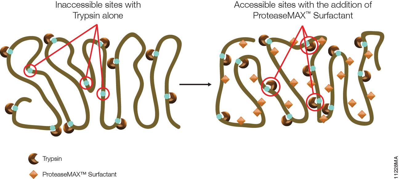
ProteaseMax 11228MA

The primary advantage of ProteaseMAX™ Surfactant is that it improves identification of proteins in gel by enhanced protein digestion, increased peptide extraction, and minimized post digestion peptide loss. However, ProteaseMAX™ Surfactant can also facilitate in-solution digestion protocols.

ProteaseMAX™ Surfactant offers two major benefits for digesting proteins in solution.

Continue reading “ProteaseMAX Surfactant: Enhanced In-solution Digestion Applications”

Rabbit Reticulocyte Lysate Translation Systems: Tools for the analysis of translational regulation

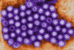
TEM of Norovirus particles. Photo Credit: Charles D. Humphrey, Centers for Disease Control and Prevention

Rabbit Reticulocyte Lysate Translation Systems are used in the identification of mRNA species, the characterization of their protein products and the investigation of transcriptional and translational control. Rabbit Reticulocyte Lysate is prepared from New Zealand white rabbits. After the reticulocytes are lysed, the extract is treated with micrococcal nuclease to destroy endogenous mRNA and thus reduce background translation to a minimum.

Untreated Lysate is prepared from New Zealand white rabbits in the same manner as treated lysates with the exception that it is not treated with micrococcal nuclease. Unlike a coupled system that initiates transcription/translation from DNA, the RNA-based rabbit reticulocyte can be used for the direct investigation of transcriptional/translational control and the replication of RNA-based viruses.


References

Characterization of translation regulation (i.e., UTRs, Capping, IRES)

  1. Nguyen, H-L .et al. (2013) Expression of a novel mRNA transcript for human microsomal epoxide hydrolase is regulated by short reading frames within it 5’ –untranslated region. RNA. 19, 752–66.
  2. Wei, J. et al. (2013) The stringency of start codon selection in the filamentous fungus Neurospora crass. J. Biol. Chem. 288, 9549–62.
  3. Paek Ki-Y. et al. (2012) Cap-Dependent translation without base-by-base scanning of an messenger ribonucleic acid. Nucl. Acid. Res. 40, 7541–51.
  4. Se, and NH. Su.W. et al. (2011) Translation, stability, and resistance to decapping of mRNA containing caps substituted in the triphosphate with BH3. RNA 17, 978–88.
  5. Anderson, D. et al. (2011) Nucleoside modifications in RNA limit activation of 2’-5’ oligoadenylate synthetase and increase resistance to cleavage by RNase L. Nucl. Acid. Res. 39, 9329-38.

RNA virus Characterization

  1. Vashist, S. et al. (2012) Identification of RNA-protein interaction networks involved in the Norovirus life cycle. J. Vir. 86, 11977–90.
  2. Soto-Rifo, R. et al. (2012) Different effects of the TAR structure on HIV-1 and HIV-2 genomics RNA translation. Nucl. Acids. Res. 40, 2653–67.
  3. Poyry, T. et al. (2011) Mechanisms governing the selection of translation initiation sites on Foot-and-Mouth Disease Virus RNA. J.Vir. 85, 10178–88.
  4. Cheng, E. et al. (2011) Characterization of the interaction between Hantavirus nucleopcapsid protein and ribosomal protein S19. J. Biol. Chem. 286, 11814–24.
  5. Vera-Otarola, J. et al. (2011) The Andes Hantavirus NSs Protein is expressed from the Viral mRMA by a leaky scanning mechanism. J. Vir. 86, 2176–87.

Compound Screening Using Cell-Free Protein Expression Systems

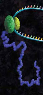
A protein chain being produced from a ribosome.
Both prokaryotic and eukaryotic cell-free protein expression systems have found great utility in efforts to screen organic compounds for inhibition of the basic cellular functions of transcription and translation, common targets for antibiotic compounds.

Cell-free systems can provide some advantages over cell-based systems for screening purposes. Cell-free systems allow exact manipulation of compound concentrations. This is an important parameter when evaluating the potential potency of the lead compound.

There is no need for cellular uptake to evaluate the effect of the compounds. While uptake evaluation is important for determining the eventual efficacy of the drug, it can unnecessarily eliminate valuable lead compounds in an initial screen. The interpretation of results in living cells is complicated by the large number of intertwined biochemical pathways and the ever-changing landscape of the growing cell. Cell-free systems allow the dissection of effects in a static system for simpler interpretation of results and the ability to specifically monitor individual processes such as transcription or translation. Individual targets not normally present, or found at low concentrations, can be added in controlled amounts.

The following references illustrate this application:

Proteinase K: An Enzyme for Everyone

protein expression purification and analysis

We recently posted a blog about Proteinase K, a serine protease that exhibits broad cleavage activity produced by the fungus Tritirachium album Limber. It cleaves peptide bonds adjacent to the carboxylic group of aliphatic and aromatic amino acids and is useful for general digestion of protein in biological samples. In that previous blog we focused on its use to remove RNase and DNase activities. However, the stability of Proteinase K in urea and SDS and its ability to digest native proteins make it useful for a variety of applications. Here we provide a brief list of peer-reviewed citations that demonstrate the use of proteinase K in DNA and RNA purification, protein digestion in FFPE tissue samples, chromatin precipitation assays, and proteinase K protection assays:

Continue reading “Proteinase K: An Enzyme for Everyone”

Protein:protein Interactions: GST Pulldowns

trypsinGST pull-downs

Pull-down assays probe interactions between a protein of interest that is expressed as a fusion protein (e.g., bait) and the potential interacting partners (prey). In a pull-down assay one protein partner is expressed as a fusion protein (e.g., bait protein) in E. coli and then immobilized using an affinity ligand specific for the fusion tag. The immobilized bait protein can then be incubated with the prey protein. The source of the prey protein can be either from a cell-based or cell-free expression system. After a series of wash steps the entire complex can be eluted from the affinity support using SDS-PAGE loading buffer or by competitive analyte elution, then evaluated by SDS-PAGE.

Continue reading “Protein:protein Interactions: GST Pulldowns”

PNGase F, a Novel Endoglycosidase

11123MA

PNGase F (Cat.# V4831) is a recombinant glycosidase cloned from Elizabethkingia meningoseptica and overexpressed in E. coli, with a molecular weight of 36kD.

PNGase F catalyzes the cleavage of N-linked oligosaccharides between the innermost GlcNAc and asparagine residues of high mannose, hybrid, and
complex oligosaccharides from N-linked glycoproteins. PNGase F will not remove oligosaccharides containing alpha-(1,3)-linked core fucose,
commonly found on plant glycoproteins.

Applications
Determining whether a protein is in fact glycosylated is the initial step in glycoprotein analysis. Polyacrylamide gel electrophoresis in the
presence of sodium dodecyl sulfate (SDS-PAGE) has become the method of choice as the final step prior to mass spec analysis. Glycosylated proteins often migrate as diffused bands by SDS-PAGE. A marked decrease in band width and change in migration position after treatment with PNGase F is considered evidence of N-linked glycosylation.

Gel based data are often correlated with information obtained from mass spec analysis. Asn-linked type glycans can be cleaved enzymatically by PNGase F yielding intact oligosaccharides and a slightly modified protein in which Asn residues at the site of de-N-glycosylation are converted to Asp, by converting the previously carbohydrate-linked asparagine into an aspartic acid, a monoisotopic mass shift of 0.9840Da is observed. The deglycosylated peptides are then analyzed by tandem mass spectrometry (MS/MS), and software algorithms are used to correlate the experimental fragmentation spectra with theoretical tandem mass spectra generated from peptides in a protein database.

Enhancing Proteomics Data Using Arg-C Protease

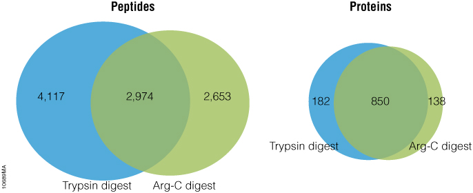
Arg-C (clostripain), Sequencing Grade (Cat.# V1881), is a specific endoproteinase isolated from the soil bacterium Clostridium histolyticum. It preferentially cleaves at the C-terminal side of arginine (R) residues. Unlike trypsin, Arg-C efficiently cleaves arginine sites followed by proline (P). This difference is important because every twentieth arginine is followed by proline. To illustrate this benefit, Arg-C was evaluated for protein analysis in two different experiments. In the first experiment, we studied the use of Arg-C for proteomic analysis. Yeast provides an excellent model proteome because its genome is well annotated. Yeast extract was digested in two parallel reactions, using trypsin in the first reaction and Arg-C in the second, using a conventional protocol consistent with LC-MS/MS analysis. As expected the trypsin digestion resulted in a high number of peptide and protein identifications (Figure 1). However, many peptides remained elusive. The parallel Arg-C digestion complemented the trypsin digestion by recovering an additional 2,653 peptides and providing a 37.4% increase in the number of identified peptides. Digesting with Arg-C also resulted in an increase in the number of identified proteins. In fact, 138 new proteins were identified in Arg-C digest compared to the parallel trypsin digest, offering a 13.4% increase in the overall number of identified proteins.

Figure 1. Side-by-side analysis of trypsin-digested and Arg-C digested yeast proteins.

Are you looking for proteases to use in your research?
Explore our portfolio of proteases today.

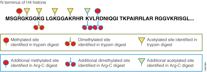
In a second experiment, the ability of Arg-C to analyze individual proteins was analyzed, selecting human histone H4 as a model protein. Like other histones, this protein is heavily modified post translational modifications (PTMs) that alter histone structure and regulate interaction with transcription factors. As a result, histone PTMs are implicated in gene regulation and associated with multiple disorders. Technical challenges, however, impede histone PTM analysis. Histone PTMs are complex and some, such as acetylation and methylation, prevent trypsin digestion, as shown by our data. In this experiment, trypsin digestion of histone H4 identified several PTMs (Figure 2). However, certain PTMs were missing. By digesting histone H4 with Arg-C, we were able to identify the missing PTMs including mono-, dimethylated and acetylated lysine and arginine residues. We speculate that the PTMs in human histone H4, which modified arginine and lysine residues, rendered trypsin unsuitable for preparing the corresponding histone regions for mass spectrometry. The problem was rectified by replacing trypsin with Arg-C.

Figure 2. Identification of histone h4 PTMs after Arg-C digestion.

Endo H Application: Monitoring Protein Trafficking

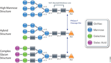
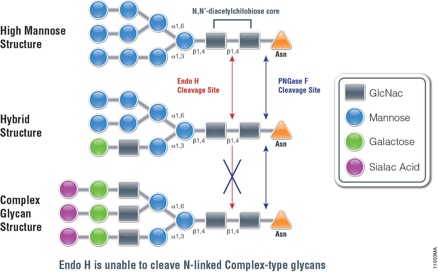
Endo H (Endo-ß-N-acetylglucosaminidase H) is a 29,000 dalton protein with optimal activity between pH 5 and 6. In contrast to PNGase F, which cleaves all N-linked glycans at the site of attachment to Asparagine (Asn), (with the exception of those with fucose attached to the core GlcNac moieties), Endo H hydrolyses the bond connecting the two GlcNac groups that comprise the chitobiose core (see Figure 1.). In addition, Endo H cleaves high mannose and hybrid glycans, whereas complex glycans (those with more than 4 different sugar types per glycan chain, including the GlcNac groups) are resistant to hydrolysis.

The unique specificty of Endo H and PNGase F can be used to monitor protein trafficking. Basic N-Glycosylation occurs in the endoplasmic reticulum. Proteins in this stage are sensitive to Endo H digestion. If proteins have entered the Golgi body where additional modifications occur to the glycan, they are resistant to Endo H digestion.

The following references illustrate this application:

Cell free application: Sumoylation characterization

Small Ubiquitin-like Modifier (or SUMO) proteins are a family of small proteins that are covalently attached to and detached from other proteins in cells to modify their function. SUMOylation is a post-translational modification involved in various cellular processes, such as nuclear-cytosolic transport,
transcriptional regulation, apoptosis, protein stability and response to stress.

In contrast to ubiquitin, SUMO is not used to tag proteins for degradation. Mature SUMO is produced when the last four amino acids of the C-terminus have been cleaved off to allow formation of an isopeptide bond between the C-terminal glycine residue of SUMO and an acceptor lysine on the target protein.

Cell free expression can be used to characterize sumoylation of proteins. Target proteins are expressed in a rabbit reticulocyte cell free system (supplemented with necessary addition components,). Proteins that have been modified can be analyzed by a shift in migration on polyacrylamide gels, when compared to control reactions.

The following references illustrate the use of cell free expression for this application.

Brandl, A. et al. (2012) Dynamically regulated sumoylation of HDAC2 controls p53 deacetylation and restricts apoptosis following genotoxic stress. J. Mol. Cell. Biol. (online only)

Janer, A. et al. (2010). SUMOylation attenuates the aggregation propensity and cellular toxicity of the polyglutamine expanded ataxin-7. Human. Mol. Gen. 19, 181—95.

Rytinki, M. et al. (2009) SUMOylation attenuates the function of PGC-1alpha. J. Biol. Chem. 284, 26184-93.

Klein, U. et al. (2009) RanBP2 and SENP3 function in a mitotic SUMO2/3 conjugation-deconjugation cycle on Borealin. Mol. Cell. Biol. 20, 410–18.

Seo, W. and Ziltener, H. (2009) CD43 processing and nuclear translocation of CD43 cytoplasmic tail are required for cell homeostasis. Blood, 114, 3567–77.