Use of Mass Spectrometry to Quantitate Food Allergens


Food allergies are becoming increasingly prevalent among children. Credit: James Gathany, CDC

Food allergies are increasing worldwide and becoming a public health issue, especially among children are concerned. Children have a higher prevalence of food allergies, with about 4–8%, compared to adults (1–5%).  Currently antibody-based methods such ELISA (enzyme-linked immunosorbent assay) are the primary method for food allergen analysis. In most cases antibodies are only available for single well-known allergens. Often those that are commercially available are poorly characterized resulting cross-reactivity that leads to false-positive results in diagnostic tests.

A recent publication (1) presented a review of an alternative technology based on mass spec (i.e., multiple reaction monitoring, MRM) that circumvents the drawbacks of antibody based methods. MRM allows precise quantitative determination of target proteins in complex samples with broad dynamic range.  MRM also provides quantification of different isoforms. It is noted that tryptic digestion followed by mass spec analysis, has already identified several unique peptides for different allergens, including those found in crustaceans, eggs, fish, peanuts, soy and wheat. In summary the challenge is now to select the appropriate tryptic signature peptide(s) for the respective allergen and to develop well characterized standards (i.e., isotope labeled standards) to ensure accurate quanititation.

Citation:

Koeberl, M et al. (2014) Next Generation of Food Allergen Quantification using Mass Spectrometric Systems J. Proteome Research  13, 3499–509.
Purifying HIS-Tagged Proteins from Insect and Mammalian Cells

MSextractcroppedMany different polypeptide fusion partners or affinity tags have been developed to facilitate purification of target proteins. The most commonly used tag for the purification and detection of recombinant expressed proteins is the His tag. Cloning vectors designed to generate His-tagged proteins contain 5–10 histidine residues at either the C- or N terminus of the expressed protein. The His tag adds only 0.84kDa to the mass of the protein and is nonimmunogenic. Also, because the tertiary structure of the tag is not important for purification, His-tagged proteins can be purified using native or denaturing conditions. The affinity of histidine residues for immobilized nickel allows selective purification of His-tagged proteins. The MagneHis™ Ni-Particles can bind up to 1mg of His-tagged protein per milliliter of particles providing a fast, efficient method for purifying His-tagged proteins with high yield and low background in a highly scalable format.
Bacterial expression of recombinant His-tagged proteins is a common technique. However, use of other systems, such as Sf9 insect cells,or HeLa or CHO mammalian cells for expression of recombinant proteins either intracellularly or secreted into the culture medium is increasing. These eukaryotic expression systems may allow more natural processing and modification of recombinant His-tagged proteins.
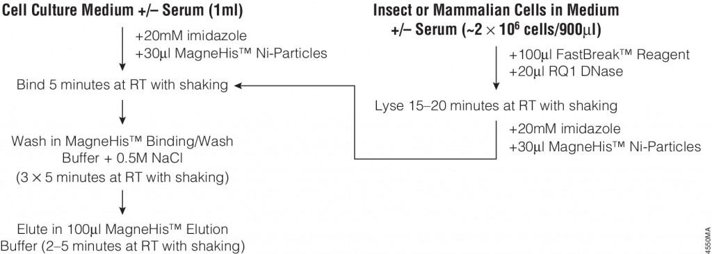
The following article:  illustrates the use of FastBreak™ Cell Lysis Reagent and the MagneHis™ Protein Purification System with insect and mammalian cell lysates. Proteins are purified from culture medium in the presence or absence of serum with only minior modifications to the standard protocol for bacterial cultures are required for purification from these diverse sources.
4550MA-(1)


Mass Spec-Compatible Proteome Reference Material

MSextractcropped

The complexity of biological samples places high demand on mass spec analytical capability. Adequate monitoring of instrument performance for proteomics studies requires equally complex reference material such as whole-cell extracts. However, whole-cell extracts available commercially are developed for general research (e.g., enzymatic or Western blot analysis) and contain detergents and salts that interfere with reverse phase liquid chromatography and mass spectrometry. Even after clean up, the extracts need to be digested, requiring time, labor and experience to generate samples for use in mass spectrometry. To address the need for complex protein material, we have developed whole-cell protein extracts from yeast and human cells. The yeast extract offers the convenience of a relatively small and well annotated proteome, whereas the human extract provides a complex proteome with large dynamic range. The human extract also serves as reference material for studies targeting the human proteome.

The extracts are free of compounds that interfere with reverse phase liquid chromatography-mass spectrometry (LC-MS), and have been reduced with DTT and alkylated with iodoacetamide then digested with Trypsin/Lys-C Mix and lyophilized. These digested extracts (tryptic peptides) can be readily reconstituted in trifluoroacetic acid (TFA) or formic acid and injected into an instrument. The same human and yeast whole-cell extracts also are provided in an intact (undigested) form for users who would like to develop an independent method for preparing protein mass spectrometry samples. For convenience, the intact extracts are provided as a frozen solution.

Consistent extract protein composition is ensured by tight control over cell culture conditions and manufacturing process. Lot-to-lot consistency of extracts is monitored by various protein and peptide qualitative and quantitation methods, including LC-MS. (Quality control results are provided upon request.) Our manufacturing process assures compatibility with reverse phase liquid chromatography and mass spectrometry, minimal nonspecific protein fragmentation, nonbiological post-translational modifi cations and,for digested extracts, minimal undigested peptides. The extracts are optimized for a high number of peptide and protein identifications in mass spectrometry analysis.

His-Tagged Fusion Proteins: Application Update

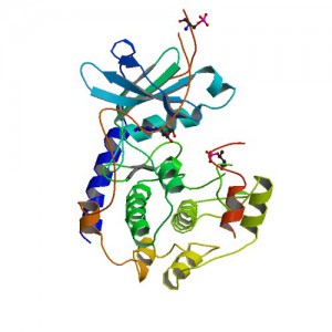
Crystal structure of Polyhistidine tagged recombinant catalytic subunit of cAMP-dependent protein kinase. Credit: StructureID=1fmo; DOI=http://dx.doi.org/10.2210/pdb1fmo/pdb;

Researchers often need to purify a single protein for further study. One method for isolating a specific protein is the use of affinity tags. Affinity purification tags can be fused to any recombinant protein of interest, allowing fast and easy purification following a procedure that is based on the affinity properties of the tag.

The most commonly used tag to purify and detect recombinant expressed proteins is the polyhistidine tag. Protein purification using polyhistidine tags relies on the affinity of histidine residues for immobilized metal such as nickel, which allows selective protein purification. The metal is immobilized to a support through complex formation with a chelate that is covalently attached to the support.

Polyhistidine tags offer several advantages for protein purification. The small size of the polyhistidine tag renders it less immunogenic than other larger tags. Therefore, the tag usually does not need to be removed for downstream applications following purification.

A large number of commercial expression vectors that contain polyhistidine are available. The polyhistidine tag may be placed on either the N- or C-terminus of the protein of interest.

And finally, the interaction of the polyhistidine tag with the metal does not depend on the tertiary structure of the tag, making it possible to purify otherwise insoluble proteins using denaturing conditions. The resulting purified protein can be used for a variety of applications.

The following references illustrate examples of some of the most common post purification applications with fusion proteins containing a polyhistidine tag:

Enzymatic assays

  1. Negi, V-S. et al. (2014) A carbon nitrogen Lyase from Leucaena leucocephala catalyzes the first Step of mimosine degradationPlant Physiol. 164,  922–34.
  2. Yu, S. et al. (2013) Syk Inhibits the activity of protein kinase A by phosphorylation tyrosine of the catalytic subunitJ. Biol. Chem. 288, 10870-81.
  3. Rusconi, B. et al. (2013) Discovery of catalases in members of the Chiamydiales order. J. Bact. 195, 3543–51.

Structural analysis

  1. Araiso, Y. et al. (2014) Crystal structure of Saccharomyces cerevisiae mitochondrial GatFAB a novel subunit assembly in tRNA –dependent amidotransferases. Nucl.Acids. Res.(available only online).
  2. Someya, T. et al. (2012) Crystal Structure of Hfq from Bacillus subtilis in complex with SELEX-dervived RNA aptamer: insight into RNA-binding properties of bacterial Hfq. Nucl. Acid Res. 40, 1856-67.

Protein pulldowns

  1. Yun, S-C. et al. (2010) Pmr a Histone-like protein H1 (H-NS) family protein encoded by the IncP-7 plasmid pCAR1, is a key global regulator that alters host functionJ.Bact. 192, 4720–31.
  2. Haim, H. et al. (2010)  Cytokeratin 8 interacts with clumping factor B: a new possible virulence factor target. Microbiology 156, 3710-21.

Additional Resources
His-tagged Protein Purification Systems

CheckMate™ Mammalian Two-Hybrid System: Application Update

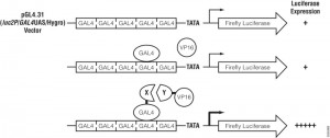
Assay principle for CheckMate™ Mammalian Two-Hybrid System.
Assay principle.

In the CheckMate™ Mammalian Two-Hybrid System, the pBIND Vector contains the yeast GAL4 DNA-binding domain upstream of a multiple cloning region, and the pACT Vector contains the herpes simplex virus VP16 activation domain upstream of a multiple cloning region. The two genes encoding the two potentially interactive proteins of interest are cloned into pBIND and pACT Vectors to generate fusion proteins with the DNA-binding domain of GAL4 and the activation domain of VP16, respectively. The pG5luc Vector contains five GAL4 binding sites upstream of a minimal TATA box, which in turn, is upstream of the firefly luciferase gene (luc+). The pGAL4 and pVP16 fusion constructs are transfected along with pG5luc Vector into mammalian cells. Interaction between the two test proteins, as GAL4 and VP16 fusion constructs, results in an increase in firefly luciferase expression over the negative controls. Traditionally mammalian two hybrid analysis was used to confirm initial data obtained from yeast two hybrid experiments.

Due to enhanced bioinformatics information and the development of improved co-immunoprecipiation/pulldown procedures/technology, there is a growing trend to use only mammalian cells to characterize protein:protein interactions. The following references illustrate the use of the CheckMate™ system to complement  other techniques to characterize protein;protein interactions using only mammalian cells .

Bagchi, P. et al. (2013) Molecular Mechanism behind Rotavirus Nsp-1 Mediated PI3 Kinase Activation: Interaction between NSP1 and the p85Subunit of PI3 kinase. J. Vir. 87, 2358-62.

Greninger, A. et al. (2013) ACBD3 Interaction with TBC1 domain 22 protein is differentially affected by Enteroviral and Kobuviral 3A protein binding.  mBio 4, 00098-13.

Patki, M. et al. ( 2013) The ETS Domain Transcription Factor ELK1 Direct a critical component of growth signalling by Androgen Receptor in prostrate cancer cells. J.Biol. Chem. 288 11047-65

Konig, H-G. (2012) Fibroblast growth factor homologous factor 1 interact with NEMO to regulate NF-kappaB signaling in neurons J. Cell Sci. 125, 6058-70

Asp-N Protease: Applications Update

TrypsinLysC Page 2

Asp-N, Sequencing Grade, is an endoproteinase that hydrolyzes peptide bonds on the N-terminal side of aspartic and cysteic acid residues: Asp and Cys. Asp-N activity is optimal in the pH range of 4.0–9.0. This sequencing grade enzyme can be used alone or in combination with trypsin or other proteases to produce protein digests for peptide mapping applications or protein identification by peptide mass fingerprinting or MS/MS spectral matching. It is suitable for  in-solution or in-gel digestion reactions.

The following references illustrate the use of Asp-N in recent publications:

Protein sequence coverage

  1. Jakobsson, M et al. (2013)  Identification and characterization of a novel Human Methyltransferase modulating Hsp70 protein function through lysine methylation. J. Biol. Chem. 288, 27752–63.
  2. Carroll, J. et. al. (2013) Post-translational modifications near the quinone binding site of mammalian complex I.  J. Biol. Chem. 288, 24799–08.

Glycoprotein analysis

  1. Siguier, B. et al. (2014) First structural insights into α-L-Arabinofuranosidases from the two GH62 Glycoside hydrolase subfamilies. J. Biol. Chem. 289, 5261–73.
  2. Vakhrushev, S. et al. (2013) Enhanced mass spectrometric mapping of the human GalNAc-type O-glycoproteome with SimpleCells. Mol. Cell. Prot. 12, 932–44.
  3. Berk, J. et al. (2013) . O-Linked β-N- Acetylglucosamine (O-GlcNAc) Regulates emerin binding to autointegration Factor (BAF) in a chromatin and Lamin B-enriched “Niche”.  J. Biol. Chem. 288, 30192–09.

Phosphoprotein analysis

  1. Roux, P. and Thibault, P. (2013) The Coming of Age of phosphoproteomics –from Large Data sets to Inference of protein Functions. Mol. Cell. Prot. 12, 3453–64.

Are you looking for proteases to use in your research?
Explore our portfolio of proteases today.

Cell-free expression application: Screening for successful oligo-mediated knockdown design

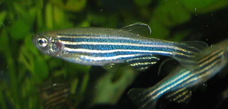
800px-ZebrafischAlthough previous references have provided data regarding the potential oncogenic role of the gene ETV7, there has been minimal investigation as to its physiological role.
In the following reference, Quintana, A. et al. (2014) Disease Models & Mechanisms 7, 265–70, zebrafish were used as in vivo model system to characterize ETV7.

One key experiment required the morpholino-oligonucleotide -mediated knockdown of in vivo ETV7. Two independent morpholinos were designed: one that inhibited translation and the other that inhibited proper splicing of exon 3. The efficacy of the translation –blocking morpholino was assessed with cell free expression of ETV7-tagged with hemagglutinin (HA).

Western blot performed with anti-HA antibodies determined the extent of the knockdown compared to a control containing no morpholino added. Once an efficient design was determined via cell-free expression screening, it was used for in vivo experiments. In conjunction additional other techniques, concluded that ETV7 is essential for normal red blood cell development.

Novel Application for ProteaseMAX Surfactant: Cell Lysis

ProteaseMax Surfactant


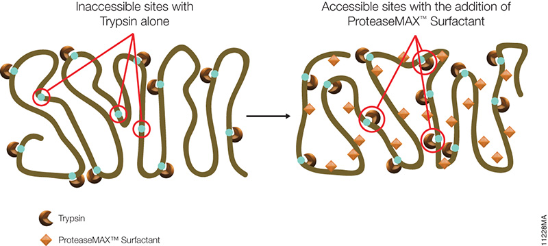
The novel mass spectrometry compatible surfactant sulfonate-(sodium 3-((1-(furan-2-yl)undecyloxy) carbonylamino)-propane-1-sulfonate (i.e.ProteaseMAX) facilitates both in-gel and in-solution digestion applications by reducing the time required, enabling protein solubilization/denaturation and increasing peptide/protein identifications.

A new application was highlighted in a recent publication (1) which utilized ProteaseMAX to lyse cells prior to trypsin digestion and subsequent mass spec analysis. The composition of the buffer determines the overall efficiency of cell lysis, dissociation of protein complexes, protein solubility and ease of removal prior to LC/MS-MS analysis.

Are you looking for proteases to use in your research?
Explore our portfolio of proteases today.

When compared to lysis buffers containing either urea or SDC, ProteaseMAX provided the optimal number of identified peptides/proteins.
In addition it can be easily removed from the lysate by acidic precipitation.

Reference

  1. Pirmoradian, M. et al. (2013). Rapid and deep human proteome analysis by single-dimension shotgun proteomics. Mol. Cell. Prot. 12, 3330–8.

ProTEV Protease Compound Compatibility Analysis

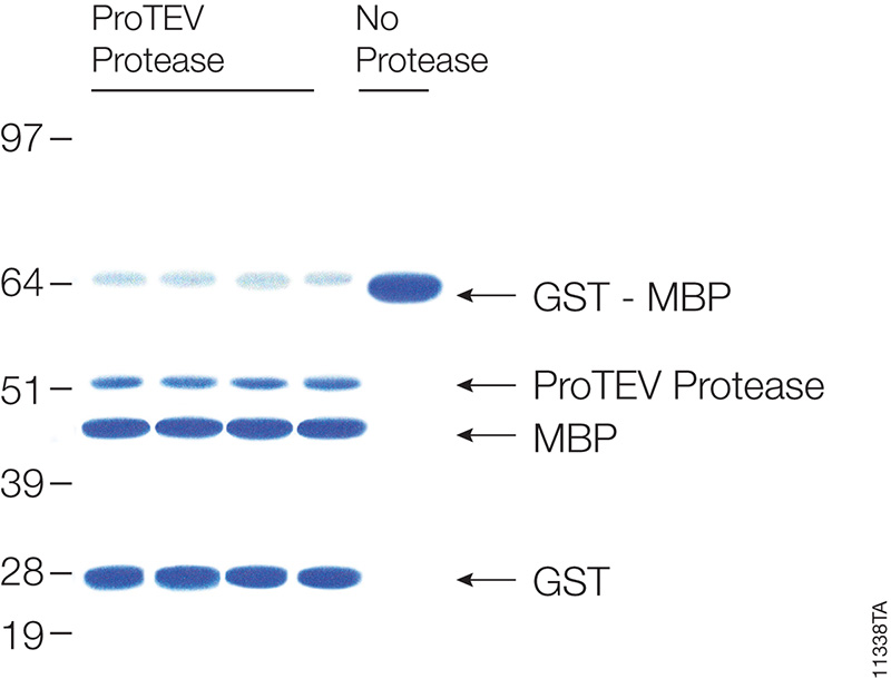
Cleavage of 20µg of GST-MBP fusion protein with ProTEV protease after 60 minutes at 30°C.

Many proteins are expressed as fusion partners with affinity tags, such as the HaloTag® fusion, glutathione-S-transferase (GST) or maltose binding protein (MBP), to selectively bind the proteins using affinity purification resins. While such resins yield high-purity protein quickly, the large affinity tags are undesirable for some downstream applications. Most expression vectors are designed with a specific protein cleavage site between the two fusion partners to remove the affinity tag after purification. ProTEV Protease recognizes a rare amino acid sequence, EXXYXQ, where X is any amino acid, and cleaves after the glutamine residue.

ProTEV Plus functions over a broad pH and temperature range. In a recent study the enzymatic activity of ProTEV Plus in the presence of various compounds (Table 1) commonly found in protein purification protocols were evaluated.

Continue reading “ProTEV Protease Compound Compatibility Analysis”

Trypsin/Lys-C Mix: Alternative for standard trypsin protein digestions

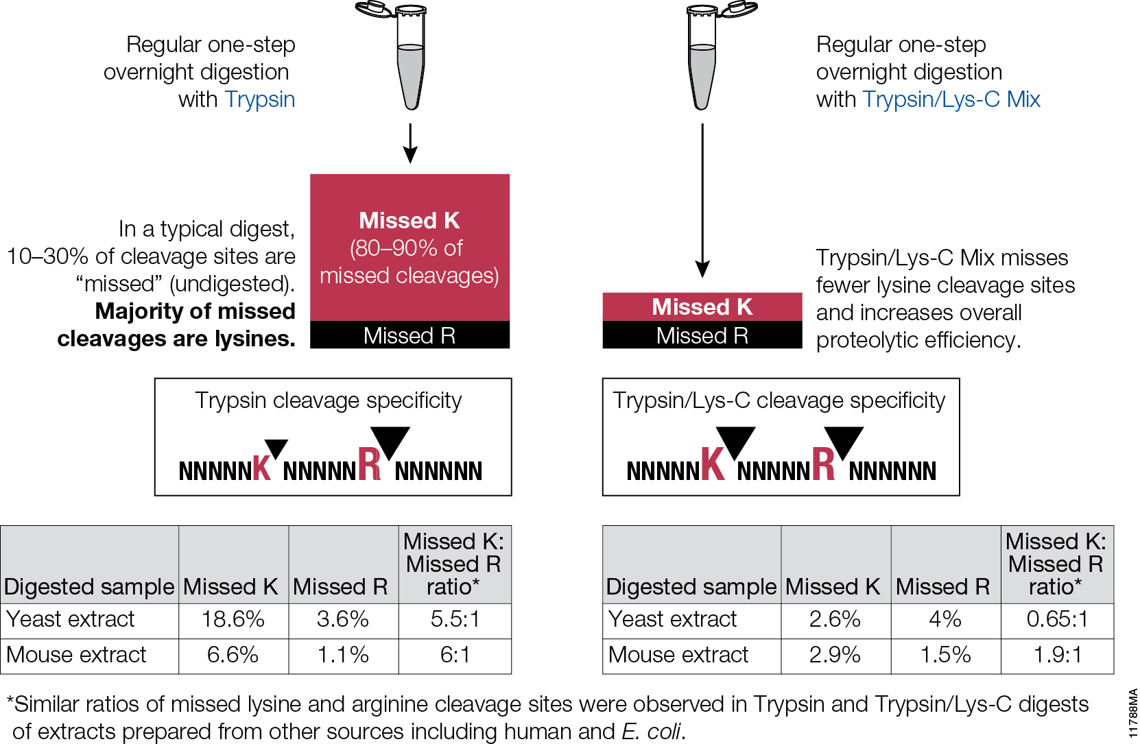
Trypsin/Lys-C Mix, Mass Spec Grade, is a mixture of Trypsin Gold, Mass Spectrometry Grade, and rLys-C, Mass Spec Grade. The Trypsin/Lys-C Mix is designed to improve digestion of proteins or protein mixtures in solution.It is a little known fact that trypsin cleaves at lysine residues with lesser efficiency than at arginine residues. Inefficient proteolysis at lysine residues is the major cause of missed (undigested) cleavages in trypsin digests.

11788MA


Supplementing trypsin with Lys-C enables cleavage at lysines with excepetional efficiency and specificity. Following the conventional trypsin digestion protocol (i.e., overnight incubation at nondenaturing conditions, reduction,alkylation, 25:1 protein:protease ratio [w/w], mix and incubate overnight at 37°C.) Replacing trypsin with Trypsin/Lys-C Mix in this conventional protocol leads to multiple benefits for protein analysis including more accurate mass spectrometry-based protein quantitation and improved protein mass spectrometry analytical reproducibility.

Are you looking for proteases to use in your research?
Explore our portfolio of proteases today.