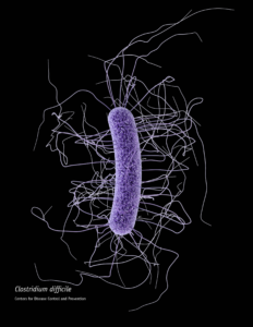
 When someone is admitted to a hospital for an illness, the hope is that medical care and treatment will help them them feel better. However, nosocomial infections—infections acquired in a health-care setting—are becoming more prevalent and are associated with an increased mortality rate worldwide. This is largely due to the misuse of antibiotics, allowing some bacteria to become resistant. Furthermore, when an antibiotic wipes out the “good” bacteria that comprise the human microbiome, it leaves a patient vulnerable to opportunistic infections that take advantage of disruptions to the gut microbiota.
One such bacteria, Clostridium difficile, is of growing concern world-wide since it is resistant to many different antibiotics. When a patient is treated with an antibiotic, C. difficile can thrive in the intestinal tract without other bacteria populating the gut. C. difficile infection is the leading cause of antibiotic-associated diarrhea. While symptoms can be mild, aggressive infection can lead to pseudomembranous colitis—a severe inflammation of the colon which can be life-threatening.
C. difficile causes disease by releasing two large toxins, TcdA and TcdB. Understanding the role these toxins play in colonic disease is important for treatment strategies. However, most published research data only report the effects of the toxins independently. A 2016 study demonstrated a method of comparing the toxins side-by-side using the same time points and cell assays to investigate the role each toxin plays in the cell death that leads to disease of the colon.
Chumbler et al. discovered that the toxins induce cell death in a concentration-dependent manner. TcdA toxin caused apoptotic cell death via glucosyltransferase activity at all concentration levels. TcdB exposure only caused apoptosis at low concentrations of toxin, but when cells were treated with a high concentration, the TcdB toxin instead induced necrotic cell death by reactive oxygen species (ROS) production.
To make these observations, the researchers developed a model using young adult mouse colonic (YAMC) epithelial cells. These cells express a temperature-sensitive version of simian virus 40 (SV40) T antigen—an oncoprotein that suppresses p53. When cultured at 33°C, p53 is suppressed and the cells are anti-apoptotic. When the temperature conditions rise to 37°C, p53 is activated and the YAMC cells undergo normal apoptosis.
With this cell culture model, Chumbler et al. measured cell viability in the presence of TcdA and TcdB toxins using the CellTiter Glo® Luminescent Cell Viability Assay. At 33°C (p53 suppressed), they found that TcdA did not cause cell death. TcdB-treated cells survived at low concentrations, but were killed at high concentrations ≥0.1nM of the toxin, suggesting that cell death at high concentrations of TcdB was not driven by apoptosis. When the cell culture conditions changed to 37°C (p53 activated), both TcdA and TcdB caused cell death at all concentrations, suggesting both toxins utilized apoptosis.
High concentrations of TcdB also induced cytotoxicity faster than TcdA. Using the CytoTox™ Glo Assay, the researchers treated the YAMC cells with concentrations of TcdA and TcdB and assessed cell membrane integrity over time at 2h, 4h, 8h, and 24h intervals. TcdB treatment caused necrotic cytotoxicity as early as 2h and up to 8h at concentrations ≥0.1nM. Low concentrations of TcdB—along with the range of TcdA treatment—only caused cell death at the 24h time point.
Finally, Chumbler et al. further investigated the mechanism of apoptotic cell death using the Apo-ONE™ Homogeneous Caspase-3/7 Assay in response to TcdA and TcdB intoxication. Caspase-3/7 activation increased in a concentration-dependent response to TcdA treatment. Low concentrations of TcdB showed caspase-3/7 activity, but apoptotic cell death was not observed at high concentrations ≥0.1nM. Along with the cell viability and cytotoxicity data, this research demonstrates that TcdB toxicity can switch between apoptotic or necrotic cell death at different concentrations.
Further understanding of how these toxins work mechanistically can help inform the development of treatments to prevent disease. A vaccine or antibody-based therapy that targets these toxins can provide immunity from infection. Research into therapies that utilize non-pathogenic strains of C. difficile bacteria also has potential. Currently, the best way to treat a C. difficile infection is by using antibiotics—only time will tell if a better, more targeted treatment becomes available before circulating strains of C. difficile become super resistant.
Learn more about Cell Health Assays.
Reference:
Chumbler, N.M. et al. (2016) Clostridium difficile toxins TcdA and TcdB cause colonic tissue damage by distinct mechanisms. Infect Immun. 84(10): 2871-7. doi: 10.1128/IAI.00583-16.
Image Source:
Centers for Disease Control and Prevention Public Health Image Library
Latest posts by Mariel Mohns (see all)
- Sustainability Makeover: Parking Ramp Edition - January 27, 2020
- Go with Your Gut: Understanding How the Microbiome and Diet Influence Health - November 26, 2019
- Kyle Hill Explains the Science of Star Wars at the Wisconsin Science Festival - October 23, 2019
