Since the 1980s, we’ve been told that aging can be accelerated by a build-up of free radicals in our cells. We’ve learned that to counteract the damage that free radicals (or reactive oxygen species, ROS) can wreak on our bodies, we should consume antioxidants like vitamins C and E, and phytochemicals.
In fact, the term “superfood” was coined for foods that contain high levels of antioxidants, phytochemicals and vitamins, foods like blueberries and carrots, spinach and kale, to name a few.
“Hold the phone”, as a pre-calculus professor of mine used to say. Turn off the blender and put down that shot glass of beet-carrot-lemon grass juice. This research just in: “Free Radicals Crucial to Suppressing Appetite”.
The research was published August 28, 2011 in the advanced online edition of Nature Medicine.
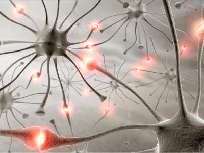
In this study, Yale University researchers reported that elevated levels of ROS in the brain activated satiety-generating neurons.
Food consumption in mice is regulated in the hypothalamus by specialized neurons. However, the neurological basis of leptin resistance, a phenomena connected to obesity in mice and humans, is unknown. Perhaps a bit of background on leptin would be helpful.
Leptin is a 16kDa protein hormone that plays a key role in regulating energy intake and energy expenditure, including appetite and metabolism.
When leptin is given to leptin-deficient ob/ob mice, it causes weight loss. However, the ability of leptin to induce weight loss in obese humans is less straightforward. Obesity can occur in the presence of high circulating levels of leptin, a phenomena known as leptin resistance.

In the current study, the authors showed that when ROS levels were suppressed, neuropeptide Y and agouti-related peptide neurons were activated, as was appetite, shown by increased feeding in mice. On the other hand, increased amounts of ROS activated pro-opiomelanocortin (POMC) neurons and reduced feeding.
In addition, levels of ROS in POMC neurons correlated with levels of leptin in lean and (ob/ob) mice, while the relationship between ROS and leptin levels was diminished in diet-induced obese mice.
The researchers induced peroxisome proliferation in POMC neurons and found ROS levels decreased, with a corresponding increase in food consumption in lean mice on a high-fat diet. However, suppression of peroxisomes by an antagonist increased free radical levels and resulted in decreased feeding in diet-induced obese mice.
The Yale group comments that their findings “unmask a previously unknown hypothalamic cellular process associated with peroxisomes and ROS in the central regulation of energy metabolism in states of leptin resistance”.
It’s amazing and humbling to learn that a molecule thought to be strictly bad for cellular health plays such an interesting feedback mechanism in the neurology of metabolism. Future work in this area could lead to interesting information both in the weight loss arena and for enthusiasts of antioxidants and superfoods.
![]() Diano S, Liu ZW, Jeong JK, Dietrich MO, Ruan HB, Kim E, Suyama S, Kelly K, Gyengesi E, Arbiser JL, Belsham DD, Sarruf DA, Schwartz MW, Bennett AM, Shanabrough M, Mobbs CV, Yang X, Gao XB, & Horvath TL (2011). Peroxisome proliferation-associated control of reactive oxygen species sets melanocortin tone and feeding in diet-induced obesity. Nature medicine, 17 (9), 1121-7 PMID: 21873987
Diano S, Liu ZW, Jeong JK, Dietrich MO, Ruan HB, Kim E, Suyama S, Kelly K, Gyengesi E, Arbiser JL, Belsham DD, Sarruf DA, Schwartz MW, Bennett AM, Shanabrough M, Mobbs CV, Yang X, Gao XB, & Horvath TL (2011). Peroxisome proliferation-associated control of reactive oxygen species sets melanocortin tone and feeding in diet-induced obesity. Nature medicine, 17 (9), 1121-7 PMID: 21873987
