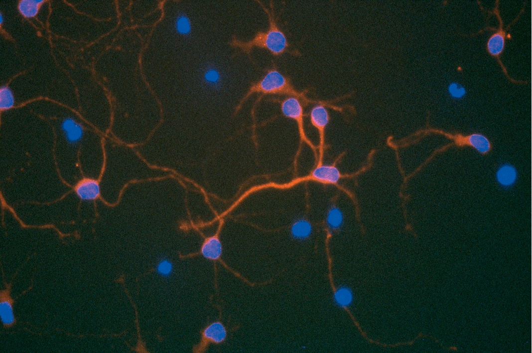
There are many levels on which science can be a beautiful thing. Some of these are quite abstract, like the experimental result that exactly proves the theory, the perfect order revealed in a mathematical equation, or the exquisite sensitivity seen in regulation of a signaling pathway. On another level the output of an experiment itself can have a physical beauty of its own. For example, immunofluorescence and immunocytochemistry technologies can generate results that not only reveal information about the subject of the experiment, but also can be quite spectacularly beautiful. Here are a few favorites of mine:
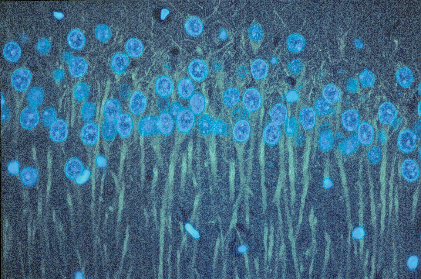
I would happily hang this on my wall. It’s a CNS section probed with an anti-beta III tubulin monoclonal antibody followed by an anti-mouse Cy3 conjugated secondary antibody, then visualized with a Cy3/Cy5 filter (yellow) and DAPI (blue).
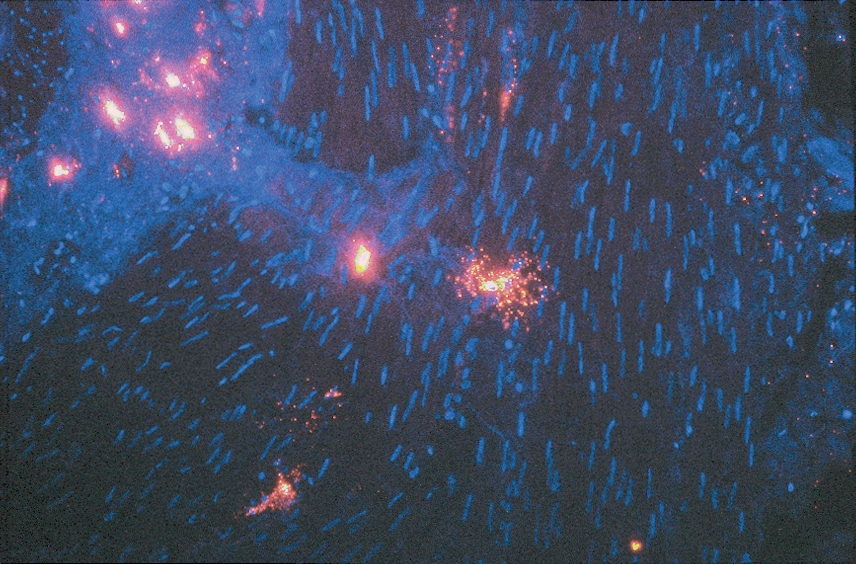
This reminds me of the famous “Starry Night”, it shows double-immunofluorescent staining of bladder cells with anti human tryptase monoclonal antibody labeled with biotin and visualized with anti-mouse Cy3 conjugate (red). Nuclei are stained with DAPI (blue).
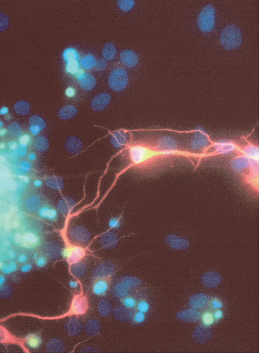
Here condensed nuclei containing fragmented DNA appear green, in contrast to the intact nuclei stained with DAPI (blue). Cells were also stained with an anti-betaIII tubulin monoclonal antibody and a CY3 labeled secondary antibody, staining immature process-bearing neurons red.
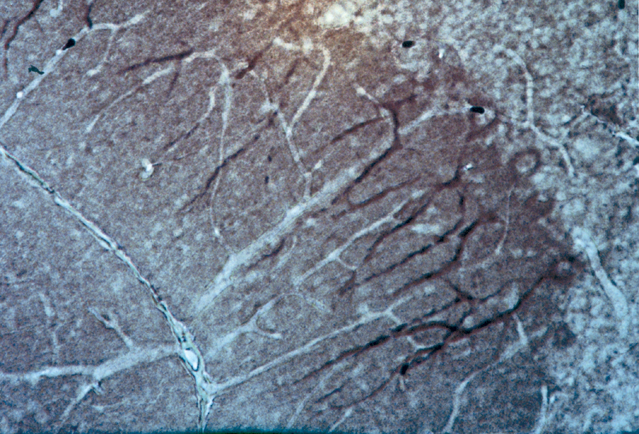
This icy looking image is actually an immunohistochemical stain of TrkB in rat cerebellum purkinje cells.
What about you? Do you have any favorite images that you would like to share?
Isobel Maciver
Latest posts by Isobel Maciver (see all)
- 3D Cell Culture Models: Challenges for Cell-Based Assays - August 12, 2021
- Measuring Changing Metabolism in Cancer Cells - May 4, 2021
- A Quick Method for A Tailing PCR Products - July 8, 2019
