Clostridium difficile is a bacterium that infects around half a million people per year in the United States. The infection causes symptoms ranging from diarrhea to severe colitis, and it’s one of the most common infections contracted while staying in the hospital. As the global incidence of C. diff infection has risen over the past decade, so has the pressure to develop novel therapeutic strategies. This requires a thorough exploration of all aspects of C. difficile biology.

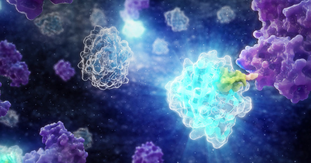
Two recent papers published by researchers at the University of Leiden have shed light on C. difficile physiology using HiBiT protein tagging. The HiBiT system allows detection of proteins in live cells using an 11 amino acid tag. The HiBiT tag binds to the complementary LgBiT polypeptide to reconstitute the luminescent NanoBiT® enzyme. The resulting luminescence is proportional to the amount of HiBiT-tagged protein that is present.
Chromatin Binding Proteins in C. Difficile
The proteins responsible for organizing and compacting nucleic acids have attracted attention as potential drug targets. The HU/IHF protein family is the most abundant class of bacterial chromatin binding proteins. HU proteins have been shown in E. coli and other bacteria to stabilized supercoiled DNA and modulate chromosome conformation. HU proteins have been shown to be essential for cell viability in Bacillus subtilis, which has one HU protein, but it is not essential in E. coli, which has three.
A 2019 paper in the Journal of Molecular Biology is the first to examine bacterial chromatin proteins in C. difficile. In this study, the authors identified the hupA gene in C. difficile, which showed significant sequence identity with homologues in E. coli, B. subtilis and S. aureus. The HupA protein also displays three conserved arginine residues that were shown to be essential for DNA binding.
Chromatography and Western blot experiments demonstrated that HupA formed homodimers in vitro, but the authors wanted to determine whether this occurred in vivo. To examine the protein interactions in vivo, the researchers genetically fused HupA to the C terminus of both subunits of a modified NanoBiT® protein. When both fusions were expressed, a luminescent signal was detected. This indicated that HupA was forming dimers, thus bringing both NanoBiT® subunits together to reconstitute the luciferase.
This study also used HaloTag to examine the localization of HupA. HaloTag® technology is based on covalent binding between the 35kDa HaloTag® protein and its various ligands. A fluorescent HaloTag® ligand can be used to visualize the location of a protein within the cell. When a HupA-HaloTag fusion was expressed in C. difficile, the localization pattern was similar to the patterns of HU proteins in other bacteria and the nucleoids appeared compressed. In contrast, when HupA was mutated to reduce its DNA binding ability, the protein was distributed throughout the cell and the nucleoid was not compacted.
Check out the complete methods and learn more about C. difficile chromatin binding proteins here:
Paiva A.M.O., et al. (2019) The Bacterial Chromatin Protein HupA Can Remodel DNA and Associates with the Nucleoid in Clostridium difficile. J Mol Biol 431, 653-72.
Regulators of Toxin Expression in C. difficile
C. difficile produces two large toxins, Toxin A and Toxin B. The locus containing these toxins (PaLoc) also encodes a TcdR, positive regulator of toxin gene expression, and TcdC, which is believed to be a negative regulator of toxin expression. TcdC is known to be a membrane-associated protein, but little is known about its topology and function.
In a 2020 article in the Journal of Bacteriology, the Nano-Glo® HiBiT Extracellular Detection System was used to determine the orientation of the C-terminus of TcdC in the membrane. The SmBiT tag was attached to the C-terminus of TcdC, and a luminescent signal was produced when LgBiT was added. Because LgBiT is too large to pass through the cell membrane, the signal indicates that the C-terminus of TcdC is located in the extracellular environment.
The HiBiT system was also used to demonstrate that a membrane-proximal cysteine residue was essential for membrane association of TcdC. When the cysteine 51 residue was mutated into serine, luciferase activity increased in the supernatant. However, total luciferase of the cells and medium was the same between the WT and mutant, indicating that the increase was not related to a change in expression.
Read the paper and learn more about the use of HiBiT for topological research in C. difficile here:
Paiva, A.M.O. et al. (2020) The C-terminal domain of Clostridioides difficile TcdC is exposed on the bacterial cell surface. J Bacteriol.
Explore more applications of HiBiT technology in these blog posts:
- CRISPR/Cas9 HiBiT Knock-In: A Scalable Approach for Studying Endogenous Protein Dynamics
- CRISPR/Cas9 Knock-In Tagging: Simplifying the Study of Endogenous Biology
Related Posts
Latest posts by Jordan Villanueva (see all)
- Tackling Undrugged Proteins with the Promega Academic Access Program - March 4, 2025
- Academic Access to Cutting-Edge Tools Fuels Macular Degeneration Discovery - December 3, 2024
- Novel Promega Enzyme Tackles Biggest Challenge in DNA Forensics - November 7, 2024

Thank you for the nice post about our use of luciferase in elucidating Cdiff physiology. Actually, I’d like to point out there is yet another paper using this: https://pubs.acs.org/doi/10.1021/acssynbio.6b00104 In this earlier manuscript we made a secreted luciferase reporter for use in Cdiff which eliminates complicated processing of samples. We have used this in subsequent work already (see for instance PMID: 30455241 PMID: 32938698). Also interesting to note that the tools we developed can be used in S. aureus too (see work from the Corrigan lab in Sheffield).