A basic tenet of immunology is that antibodies produced by B cells are very important and specific immunoprotective agents, released in response to infection.
However, antibodies do not supply immediate protection. The invading organism needs to get into the host, meet up with T cells and then B cells, in order for antibody production to occur. If the host has seen this particular pathogen previously, the antibody response occurs somewhat more quickly, but we’re still talking about days. If the invading organism is a bacterium, it can multiply and double in numbers in just hours. Thus an infection could potentially gain a foothold in a body prior to an antibody response.
Fortunately we have a more rapid, first line of defense to invading pathogens, a cellular response. In the case of a puncture or skin wound, epithelial cells, mast cells and leukocytes are activated quickly in response to pathogens. Neutrophils and monocytes also aid the cellular response.
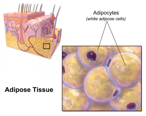
Now a recently published report demonstrates that fat cells also play a part in the cellular response to invading bacteria. R. Gallo et al. published a study on Jan. 2 in Science, providing more in depth information on the role of adipocytes in the host response to the bacterium Staphylococcus aureus (S. aureus).
S. aureus is of particular interest because it is: 1) a major cause of skin and soft-tissue infections in humans; and 2) the bacterium responsible for MRSA (methicillin-resistant Staph. aureus), a growing problem in community-acquired Staph infections and not treatable by any currently existing antibiotics. MRSA is a very dangerous bacterium, indeed. I think the following quote says it all, regarding S. aureus:
“S. aureus has evolved a comprehensive strategy to address the challenges posed by the human immune system.” G. Liu
There is previous evidence of adipocytes (fat cells) having immunological abilities. Unique to the work by Gallo et al. is the recognition that there is expansion of the subcutaneous adipose layer, in response to S. aureus infection.
These authors showed an increase in lipid staining, an increase in abundance of adipocytes and increased activation of the adiponection promoter. They also demonstrated an increase in the size of adipocytes due to S. aureus infection. Skin cells from S. aureus -infected mice showed greater adipogenic potential than that of cells isolated from noninfected skin. The authors confirmed that proliferation was associated with adipocyte formation by BrdU incorporation. A significant number of cells with BrdU-positive nuclei were also positive for caveolin, an adipocyte surface protein, proof that the differentiating cells were not myeloid in origin.
To determine whether adipocyte activation was essential, the researchers compared normal and adipogenesis-impaired mice. Results showed that adipogenesis impaired mice were more susceptible to S. aureus infection, both at the site of infection and in subsequent S. aureus bacteremia that was not detected in the controls.
Furthermore, the authors showed that adipocytes defend by the release of an antimicrobial peptide. They used a cell line that can be induced to mature into adipocytes, 3T3-L1 cells, and looked for expression of the antimicrobial peptide, cathelicidin. Comparison of preadipocyte 3T3-LT1 and mature adipocytes showed that cathelicidin was strongly induced in the mature, but not in the precursor cells. Similar studies with cultured primary human preadipocytes also showed cathelicidin production was induced during differentiation.
When 3T3-L1 cells were exposed to S. aureus-conditioned medium or to UV-killed bacteria, cathelicidin mRNA production was significantly enhanced, showing that the presence of S. aureus during adipogenesis enhances the release of antimicrobial peptide.
Finally the authors showed that mice in which adipose expansion was blocked, had reduced defense to S. aureus, and reduced cathelicidin expression.
A major point that I overlooked in first reading this paper is that it is not the presence of fat, but rather the differentiation of fat precursor cells into mature fat cells, that provides the antimicrobial peptide by which adipocytes attack S. aureus. The authors note that,
“Local expansion of dermal fat produces cathelicidin in response to infection, but this response appears to decline as adipocytes mature.“
Yet another reminder that the ability to make fat is important, while the presence of fat can be problematic.
