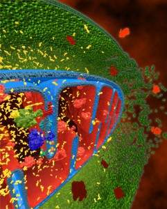
Drug-induced mitochondrial toxicity is a concern for pharmaceuticals that was, until recently, limited by the availability of a cell-based assay that is amenable to rapid high-throughput screening. Incorporating high-throughput assay chemistry that can detect mitochondrial dysfunction early in drug discovery programs provides the opportunity to identify potential mitotoxicants before they reach clinical trials or the market population.
In the paper reviewed here (1) Swiss and colleagues have validated the use of a commercially available Mitochondrial ToxGlo™ Asssay (2) to replace traditional methods that not only require expensive reagents and/or equipment but also do not lend themselves easily to high-throughput screening formats.
An additional bottleneck encountered in identifying mitochondrial toxicity is that many of the cell lines used in high-throughput drug discovery screens tend to be resistant to compounds that disrupt mitochondrial oxidative respiration when grown in glucose-containing media, a phenomenon known as the Crabtree effect. On the contrary, cells cultured in galactose-containing medium rely on their mitochondria for ATP production, and are thus more susceptible to mitochondrial inhibition (3). The authors (1) demonstrate that Mitochondrial ToxGlo™ Assay can potentially be performed in galactose-containing medium or in glucose-containing medium, provided proper validation studies are carried out initially.
The Mitochondrial ToxGlo™ assay uses a dual parameter format performed in the same well with bioluminescent and fluorescent readouts. The bioluminescent signal correlates positively with the ATP content in the cells whereas the fluorescent signal is a quantitative measure of loss of membrane integrity. The two sets of data are then combined to produce profiles representative of mitochondrial dysfunction independent of or in conjunction with other cytotoxic mechanisms. The assay can be performed with a variety of cell types including stem cell-derived cell lines; moreover, as demonstrated for K562 cells, a human immortalized myelogenous leukemia cell line, it can be completed with a short (2 h) compound incubation time.
The authors validated the dual parameter assay by testing a selection of six compounds including mitochondrial toxicants and non-mitochondrial toxicants in K562 cells, grown in both glucose- and galactose- media and showed that the assay has good reproducibility in 384-well format. They then extended the studies to test total of 74 compounds, some of which are known mitochondrial inhibitors. They defined stringent scenarios for computing the toxic effects of a test compound which depended on cellular ATP levels being depleted sooner or at lower compound concentrations than disruption of cell membrane integrity. Based on this they categorized the compounds that were tested into 4 groups; (i) those that cause mitochondrial toxicity (e.g. antimycin), (ii) those that cause multifactorial toxicity without mitochondrial impairment (e.g. digitonin), (iii) those that are nontoxic (e.g. trazodone) and (iv) those that cause multifactorial toxicity with mitochondrial dysfunction (e.g. atorvastatin). Although the compound set was tested in K562 cells in both glucose and galactose media, they show that the assay can be performed with K562 cells in galactose medium alone.
But they caution that it would not have been possible to identify mitotoxins in K562 cells if glucose medium alone had been used since the compounds in Category 1 would not have been identified as mitochondrial toxicants. They highlight the importance of initially doing a validation study with the cells of one’s choice with cell culture medium most appropriate for that cell type so that compounds that are canonical mitochondrial toxins (e.g. antimycin) can be easily distinguished from compounds that are toxic but clearly not mitochondrial toxins (e.g. digitonin).
In addition to choosing the most suitable cell culture medium when using the cell type of one’s choice, it is also necessary to choose an appropriate incubation period when testing compounds. An incubation that is too long, may incorrectly identify a mitochondrial toxin as one that causes multifactorial toxicity since prolonged incubation at lower doses in glucose-media may show cytotoxic effects similar to short incubations at higher doses in galactose-media. Conversely, an incubation time that is too short may fail to identify compounds that are toxic. For instance, a subcategory of compounds that were identified in this assay using K562 cells had no effect on either ATP levels or membrane integrity in either glucose- or galactose- medium but are known to cause mitochondrial impairment in other assays. Hence, an initial validation study should be done with a selection of compounds that (a) are classical mitochondrial inhibitors and (b) deplete ATP simply because of apoptosis/necrosis, so that an optimal incubation time is selected.
In conclusion, the assay deserves attention as a robust, early readout of mitochondrial and cellular toxicity, with companion assays being established to detect mitochondrial toxicity when lead compounds have been selected.
Reference:
- Swiss, R. et al. (2013) Validation of a HTS-amenable assay to detect drug-induced mitochondrial toxicity in the absence and presence of cell death. Toxicology in Vitro 27, 1789–1797.
- Mitochondrial ToxGlo™ Assay Technical Manual #TM357, Promega Corporation.
- Arduengo, M. Differentiating Mitochondrial Toxicity from Other Types of Mechanistic Toxicity. [Internet] May 2012. [cited: year, month, date]. Available from: http://www.promega.com/resources/pubhub/differentiating-mitochondrial-toxicity-from-other-types-of-mechanistictoxicity/
History
Updated April 2024 to correct broken link.
Radhika Ganeshan
Latest posts by Radhika Ganeshan (see all)
- T-Vector Cloning: Answers to Frequently Asked Questions - February 19, 2016
- Beating the Odds of Cancer: Not Just a Tall Tale - October 12, 2015
- Removing Cancer’s Cloak of Invisibility - April 20, 2015
