 I am fascinated by all the ways that scientists are taking sensitive techniques and using them to look into our past. For example, scientists constructed the entire genome of Yersinia pestis, the caustive agent of the Black Death, from teeth and bone samples of plague victims from the 14th century. Without methods like polymerase chain reaction (PCR), such an analysis could not be performed. My fellow blogger Terri discussed how a postmortem autopsy of Ozti, a mummy found in the Alps, used modern techniques to learn not only what color his eyes were but that he suffered from Lyme disease. In a recent PLOS ONE article, Corthals et al. took this analysis of preserved human remains further to determine if a mummy from the Andes in Argentina may have suffered from an active lung infection, testing for an immune response by protein profiling.
There were three 500-year-old Andean mummies found in 1999, all remarkably well preserved. Scans had shown the 15-year-old girl, nicknamed the Maiden, had a upper respiratory infection, while the 7-year-old boy (simply called the Boy) had no markers of infection on scans, making him the control profile for this experiment. Obtaining the samples for analysis was simple and not invasive: swabbing the lips of the Maiden and the Boy and taking a small sample of blood-soaked fabric from the Boy. These samples were processed to purify the protein fragments or peptides. Even under the best preservation conditions, proteins break apart over time, producing peptides. The Maiden’s and Boy’s purified peptides were then analyzed using mass spectrometry, a method that separates fragments by size and can be used for sequencing proteins. The mass spectrometry profiles were compared to a database of protein mass spectra to see if any matches could be identified.
While the recovered peptide profiles yielded a host of matching proteins in the database, the scientists were primarily interested in proteins involved in inflammatory and immune response. In the Maiden there was clear evidence of an immune response linked to a respiratory infection. A protein hallmark of inflammation and neutrophil infiltration of the airways, cathepsin G, was found. They also detected α-1 antitrypsin, a strong indicator of mycobacterial infection and associated with chronic lung inflammatory diseases. Proteins associated with macrophage response to lung tissue inflammation and groups of proteins that indicate severe lung inflammation were present in the Maiden’s samples. However, the Boy’s samples did not have the same protein markers for lung inflammation, although the Boy and the Maiden did share other proteins not associated with inflammation. It was only the respiratory infection markers that differed.
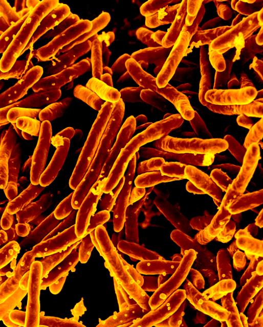
Some of the proteins that indicated severe lung inflammation are associated with acute mycobacterial infections of the lung. DNA was purified from swab samples using two different methods after extensive pretreatment of the amplification area (UV treatment and cleaning with bleach) to prevent contamination of the 500-year-old DNA samples with modern DNA. Mock DNA extractions and blank (no DNA template) PCRs were used as controls to test for any contamination. The Maiden’s and Boy’s DNA were amplified using primers specific for the genus Mycobacterium as well as specific to Mycobacterium avium and Mycobacterium tuberculosis, the latter two bacteria associated with pathogenic infections in humans, and PCR products were sequenced to compare to known sequences from this genus and species. The Boy’s DNA did not amplify any fragments, the Maiden’s DNA was positive for all amplified regions and the controls showed no amplification. Interestingly, another genus of bacteria associated with the gastrointestinal tract was amplified from the Maiden and Corthals et al. hypothesized that its presence was not contamination but indicated that the Maiden vomited prior to death.
This study used protein analysis by mass spectrometry with PCR and sequencing analysis to examine the immune response and confirm an active infection prior to death. This shotgun proteomics technique as named by the authors is a more sensitive method that can be used to detect proteins in samples and improve diagnosis of an infectious agent (e.g., Mycobacterium) in context (active infection versus passive latency). The authors speculate about its uses in forensics and other historical analysis. However, the analysis performed in the PLOS ONE article was dependent on having access to a large protein database of mass spectrometry profiles and a second sample of a putatively healthy individual from the same time period in the same location preserved under the same conditions as a control. This technique has promise, and I hope we will soon see more scientists using this analysis for their research.
Reference
Corthals A, Koller, A., Martin, D.W., Rieger, R., Chen, E.I., Bernaski, M., Recagno, G. and Dávalos, L.M. (2012). Detecting the immune system response of a 500 year-old inca mummy. PLOS ONE, 7 (7) PMID: 22848450
Sara Klink
Latest posts by Sara Klink (see all)
- A One-Two Punch to Knock Out HIV - September 28, 2021
- Toxicity Studies in Organoid Models: Developing an Alternative to Animal Testing - June 10, 2021
- Herd Immunity: What the Flock Are You Talking About? - May 10, 2021
