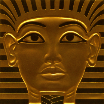
The pharaoh Tutankhamun ruled ancient Egypt during the 18th dynasty of the New Kingdom (circa 1550–1295 BC), one of the most powerful royal houses in ancient Egypt. Although he sat on the throne for only 9 years and died at the young age of 19, he is one of the most well known pharaohs, due largely in part to the discovery of his intact tomb by archaeologist Howard Carter in 1922. The tomb was filled with riches befitting a king, including an elaborate sarcophagus with a gold burial mask and statues of ancient Egyptian gods such as Osiris and Anubis, as well as gold jewelry, statues and images of servants, ornate furniture, models of boats and other items that the pharaoh would need in the afterlife. A lesser known item discovered in his tomb was an undecorated wooden box in which two small gilded coffins lay side by side. These coffins held the mummified remains of King Tutankhamun’s two stillborn daughters. Recently, researchers examined these remains in detail to determine their gestational ages and characterize any congenital abnormalities that they might have inherited from the boy king.
The two tiny mummies were first examined in 1925, when Carter unwrapped the mummies for closer inspection, then again in 1932 when a professor at Cairo University performed more complete autopsies. At that time, both mummies were well preserved. The larger mummy even had eyebrows, eyelashes and fine hair on its head. The mummies were placed in storage until 1978, when radiographic examinations were performed on the larger mummy. Based on their analysis, the researchers concluded that the fetus had a gestational age of at least 35 weeks but could have been near full term. They also reported multiple congenital abnormalities of the scapula and spine, including neural tube defects and scoliosis.
However, both mummies had deteriorated considerably since their discovery in 1922. Could postmortem damage have been interpreted as skeletal anomalies?
In November of 2011, researchers at Cairo University and the Department of State for Antiquities published their results of multi-detector computerized tomography (MDCT) analysis of both mummies (1). This advanced imaging technology generates higher-resolution images than radiographic imaging, allowing researchers to see details that were not discernible in 1978.
These images revealed just how much damage these mummies had sustained. The larger mummy was in better shape than the smaller mummy, but both exhibited extensive skull fractures, making it impossible to take measurements and assess the degree of cranial suture fusion to determine gestational age. Instead, the degree of ossification and the length of the humerus bones were used to estimate their ages as 25 weeks for the smaller mummy and 37 weeks for the larger mummy. The larger mummy also showed multiple postmortem fractures of the spine, shoulders, arms and feet.
The smaller mummy showed no obvious signs of skeletal abnormalities, although the mummy was in very poor condition by that time. Images of the larger mummy clearly showed that the left shoulder was displaced upward and the clavicle was dislocated and that this damage occurred after mummification, indicating that the fetus did not have a shoulder deformity. Likewise, the alleged defects of the skull, arms, wrists and feet were attributed to postmortem damage, not inherited defects. In fact, the only evidence of true malformations was a slight curvature of the spine. These high-resolution images gave no clues as to the cause of death for either fetus.
Considering what we now know about King Tutankhamun’s ancestry, the presence of skeletal anomalies would not be surprising. In 2010, a team of researchers performed STR and radiologic analyses of 11 royal mummies (2) from the New Kingdom to determine their genetic relationships and characterize any pathologies. These analyses allowed researchers to develop a royal pedigree and confirm that Tutankhamun was the father of the two stillborn girls. They also showed that King Tutankhamun’s parents, grandparents and great-grandmother exhibited many signs of bone abnormalities, including scoliosis, kyphoscoliosis, clubfoot and cleft palate. King Tutankhamun was the product of a consanguineous marriage between the pharaoh Akhenaten and one of his five sisters, and he suffered for it. Radiographic examinations of Tutankhamun’s mummy revealed that he had a cleft palate, mild clubfoot of the left foot and mild kyphoscoliosis and that the middle phalanx of one of his toes was absent (2). He also suffered from Köhler disease II, a rare bone disorder where a loss of blood supply causes necrosis and deterioration of a specific bone in the foot. As a result, Tutankhamun had trouble walking: In many images made during his reign, he is depicted as sitting while engaged in activities where normally he would be standing, and his tomb contained 130 walking sticks. Tutankhamun’s wife, and the mother of the two fetuses, might also be his half-sister, further increasing the risk that his two daughters would inherit one of his many maladies.
Despite these risks, King Tutankhamun’s daughters showed very few signs of malformations, and we are still left to guess as to the cause of death. Even with our advanced technology, we may never know why his two daughters were stillborn, leaving the boy king with no heirs to the throne.
References
- Hawass, Z. and Saleem, S.N. (2011). Mummified daughters of King Tutankhamun: Archaeologic and CT studies. Am. J. Roentgenol. 197, W829–36. PMID 22021529
- Hawass, Z. et al. (2010) Ancestry and pathology in King Tutankhamun’s family. JAMA 303, 638–47. PMID 20159872


Well written.
Thank you Dr. Saleem. I hope you enjoyed reading the article as much as I enjoyed writing it.